. 2009 Dec 8:9:144.
doi: 10.1186/1471-2229-9-144.

Cytosolic N-terminal arginine-based signals together with a luminal signal target a type II membrane protein to the plant ER

Affiliations

Cytosolic N-terminal arginine-based signals together with a luminal signal target a type II membrane protein to the plant ER

Aurélia Boulaflous et al. BMC Plant Biol. .

Abstract

Background: In eukaryotic cells, the membrane compartments that constitute the exocytic pathway are traversed by a constant flow of lipids and proteins. This is particularly true for the endoplasmic reticulum (ER), the main "gateway of the secretory pathway", where biosynthesis of sterols, lipids, membrane-bound and soluble proteins, and glycoproteins occurs. Maintenance of the resident proteins in this compartment implies they have to be distinguished from the secretory cargo. To this end, they must possess specific ER localization determinants to prevent their exit from the ER, and/or to interact with receptors responsible for their retrieval from the Golgi apparatus. Very few information is available about the signal(s) involved in the retention of membrane type II protein in the ER but it is generally accepted that sorting of ER type II cargo membrane proteins depends on motifs mainly located in their cytosolic tails.

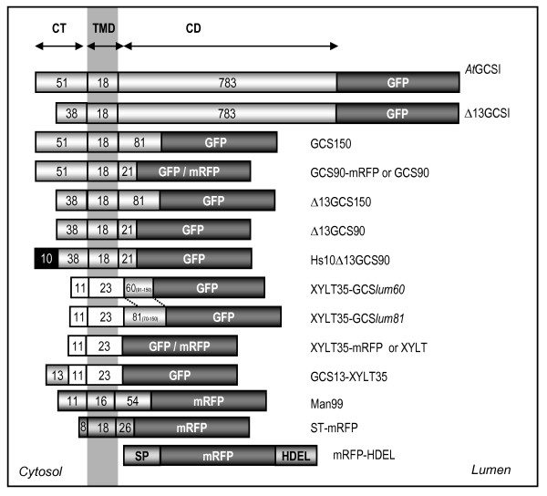
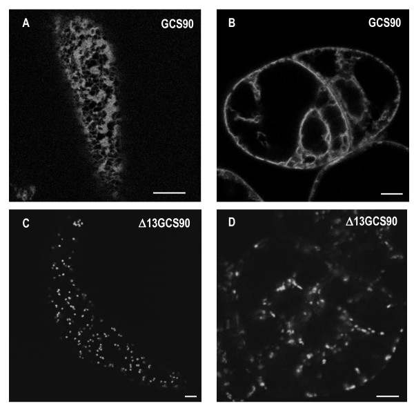
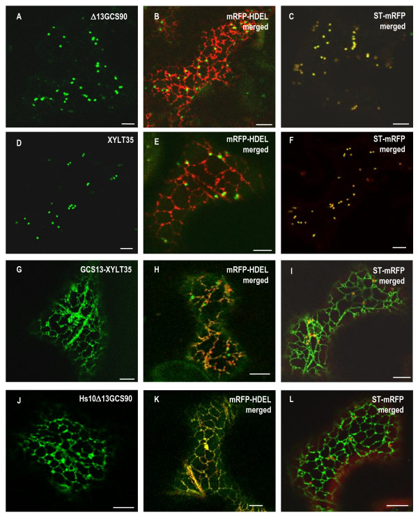
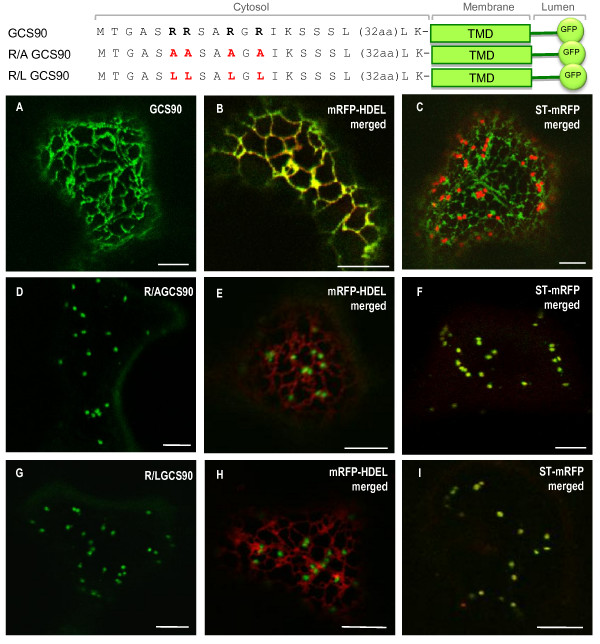
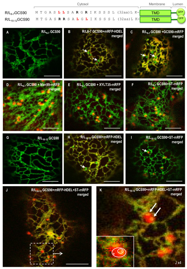
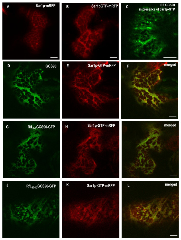
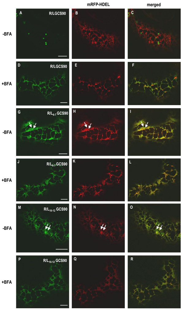
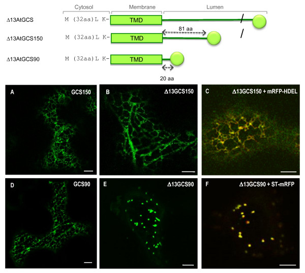
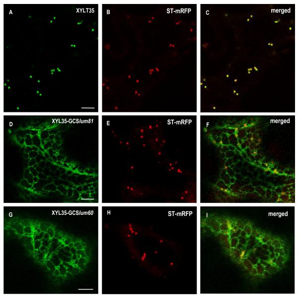
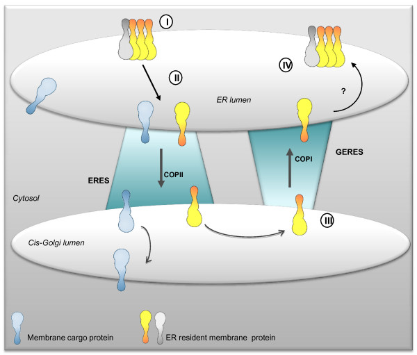
Results: Here, using Arabidopsis glucosidase I as a model, we have identified two types of signals sufficient for the location of a type II membrane protein in the ER. A first signal is located in the luminal domain, while a second signal corresponds to a short amino acid sequence located in the cytosolic tail of the membrane protein. The cytosolic tail contains at its N-terminal end four arginine residues constitutive of three di-arginine motifs (RR, RXR or RXXR) independently sufficient to confer ER localization. Interestingly, when only one di-arginine motif is present, fusion proteins are located both in the ER and in mobile punctate structures, distinct but close to Golgi bodies. Soluble and membrane ER protein markers are excluded from these punctate structures, which also do not colocalize with an ER-exit-site marker. It is hypothesized they correspond to sites involved in Golgi to ER retrotransport.

Conclusion: Altogether, these results clearly show that cytosolic and luminal signals responsible for ER retention could coexist in a same type II membrane protein. These data also suggest that both retrieval and retention mechanisms govern protein residency in the ER membrane. We hypothesized that mobile punctate structures not yet described at the ER/Golgi interface and tentatively named GERES, could be involved in retrieval mechanisms from the Golgi to the ER.


Figures

Figure 1
Figure 1
Schematic representation of the fusion proteins analyzed in this study. AtGCSI: full-length A. thaliana α-glucosidase I fused to GFP. Δ13GCSI: GCSI minus the first 13 N-terminal aa (MTGASRRSARGRI-). GCS150: the first 150 aa of GCSI fused to GFP. GCS90: the first 90 aa of GCSI fused to GFP or mRFP. Δ13GCS150: GCS150 minus the first 13 N-terminal aa. Δ13GCS90: GCS90 minus the first 13 N-terminal aa. Hs10-Δ13GCS90: the first 10 N-terminal aa of Homo sapiens GCSI (MARGERRRRA-) fused at the N-terminus of Δ13GCS90. XYLT35: the first 35 aa of A. thaliana β-1,2-xylosyltransferase fused to GFP or mRFP [47]. XYLT35-GCSlum60: the first 35 aa of XYLT fused to the first 60 aa of the luminal domain of GCSI (Pro91 to Cys150) and to GFP. XYLT35-GCSlum81: the first 35 aa of XYLT fused to the first 81 aa of luminal domain of GCSI (Arg70 to Cys150) and to GFP. GCS13-XYLT35: the 13 first N-terminal aa of GCSI fused to XYLT35. ST-mRFP: the first 52 aa of a rat α-2,6-sialyltransferase (ST) fused to mRFP [90]. mRFP-HDEL: mRFP under the control of the sporamine signal peptide and the HDEL ER retention sequence. CT: cytosolic tail; TMD: transmembrane domain; CD: C-terminal domain.
Figure 2
Figure 2
The 13 first N-terminal amino acids of AtGCSI contain ER targeting information. CLSM analysis of transgenic tobacco BY-2 cells showing cortical views (A, C) or cross sections (B, D). (A, B) GCS90 accumulates in the ER in BY-2 suspension-cultured cells. (C, D) Δ13GCS90 accumulates into the Golgi apparatus. Bars = 8 μm.
Figure 3
Figure 3
Arginine-rich ER targeting sequences are conserved for GCSI between kingdoms. CLSM analysis of Nicotiana tabacum leaf epidermal cells expressing GFP fusions alone (left panels), or co-expressing GFP fusions and either the ER marker mRFP-HDEL (middle panels), or the Golgi marker ST-mRFP (right panels). Δ13GCS90 (A-C) is exclusively located in the Golgi and perfectly co-localizes with ST-mRFP (C). XYLT35 is also located in the Golgi (D-F); [44]. When GCS13-XYLT35 (G) is co-expressed with mRFP-HDEL, the ER appears in yellow and the Golgi remains green (H) whereas when GCS13-XYLT35 is co-expressed with ST-mRFP the Golgi is yellow and the ER is green (I) showing GCS13-XYLT35 has a dual location in the ER and in the Golgi. Interestingly, when the first 13 N-terminal amino acids of GCS90 are replaced by the first 10 N-terminal amino acids of the human GCSI, Hs10Δ13GCS90 is located exclusively in the ER (J) as illustrated from colocalization with mRFP-HDEL (K) and the absence of overlap for GFP and RFP signals when it is co-expressed with ST-mRFP (L). This together with data presented Table 1 suggests that arginine-rich ER targeting sequences are conserved for GCSI between kingdoms. Bars = 8 μm.
Figure 4
Figure 4
The N-terminal arginine residues of AtGCSI contain ER localization information. CLSM analysis of Nicotiana tabacum leaf epidermal cells expressing GFP fusions alone (left panels), or co-expressing GFP fusions and the ER marker mRFP-HDEL (middle panels), or co-expressing GFP fusions together with the Golgi marker ST-mRFP (right panels). GCS90 (A) co-localizes with mRFP-HDEL (B, ER in yellow) but not with ST-mRFP (C, ER in green, Golgi in red). When the four arginine residues in position 6, 7, 10 and 12 are replaced by alanine or leucine residues, R/A GCS90 (D-F) or R/L GCS90 (G-I) accumulates exclusively in the Golgi showing that arginine residues are involved in AtGCSI ER localization. Bars = 8 μm.
Figure 5
Figure 5
Punctate structures do not accumulate ER resident proteins and are distinct from Golgi stacks. When arginine residues are mutated by pairs, R/L6-7GCS90 (A-I) and R/L10-12GCS90 (G-K) are located in the ER (A, G). Co-expression with soluble ER marker mRFP-HDEL (B, H) or membrane (C) ER marker GCS90-mRFP reveals those markers are excluded from the punctate structures that appear in green (arrows). Punctate structures are closely associated to Golgi stacks labelled with the cis-Golgi marker Man99-mRFP (D), the medial Golgi marker XYLT35-mRFP (E) or trans-Golgi marker ST-mRFP (F, I). When the constructs highlighting punctate structures are co-expressed together with the ER marker mRFP-HDEL and the Golgi marker ST-mRFP, the ER and the punctate structures appear in yellow (J). When zooming, micrograph suggests punctate structures can be closed to the ER (K, top and bottom arrows). Zone I corresponds to the co-localization area between a punctate structure and a Golgi whereas zone II corresponds to the Golgi only (K, insert). Arrows indicate the punctate structures.
Figure 6
Figure 6
Sar1p-GTP regulates ER to Golgi traffic of GCS90 and induces the disappearance of the punctate structures. CLSM analysis of Nicotiana tabacum leaf epidermal cells expressing GFP-fusions simultaneously with Sar1p variants. Sar1p-mRFP (A) and Sar1p-GTP-mRFP (B) are accumulated at the ER. Because Sar1p-GTP-mRFP blocks ER exit, membrane proteins accumulate in the ER and the ER membrane morphology turns into fenestrated sheets (B). In presence of Sar1p-GTP-mRFP, the Golgi fusion R/LGCS90 is blocked in the ER (C, compare with pattern presented Figure 5G). When GCS90 (D-F), R/L6-7GCS90 (G-I) or R/L10-12GCS90(J-L) are co-expressed with Sar1p-GTP-mRFP, the expression patterns remain unchanged, (compare to Figure 7G-I, A-C and D-F, respectively), except that the punctate structures have disappeared. Bars = 8 μm.
Figure 7
Figure 7
Punctate structures diappear when COPI-mediated retrograde transport is inhibited with BFA. CLSM analysis of Nicotiana tabacum leaf epidermal cells co-expressing GFP-fusions and mRFP-HDEL. Control cells co-expressing R/LGCS90 and mRFP-HDEL show green Golgi stacks and a red ER (-BFA, panels A-C). When cells are treated with BFA for 2 h, Golgi membranes are reabsorbed in the ER and the ER appears in yellow (+BFA, panels D-F). When cells co-expressing R/L6-7GCS90 (G-L) or R/L10-12GCS90 (M-R) and mRFP-HDEL are treated with BFA for 2 h, punctate structures disappear (J-L and P-R respectively).
Figure 8
Figure 8
The N-terminal arginine motifs are not the unique determinants responsible for ER retention of AtGCSI. When expressed in Nicotiana tabacum leaf epidermal cells Δ13GCS150-GFP is located in the ER (B) A as it was observed for the GCS150 (A) and confirmed after co-expression with the ER marker mRFP-HDEL (C). In contrast, Δ13GCS90-GFP is targeted to the Golgi apparatus (E) where it colocalizes with the Golgi marker ST-mRFP (D) whereas GCS90 is accumulated in the ER (D). Bars: 8 μm.
Figure 9
Figure 9
A luminal domain of AtGCSI is sufficient for targeting a Golgi marker into the ER. (A-C) XYLT35 and ST-mRFP are targeted to the Golgi when expressed in Nicotiana tabacum leaf epidermal cells. (D-F, G-I) XYLT35-GCSlum81 or XYLT35-GCSlum60 where co-expressed with ST-mRFP. Both fusion proteins containing a 81 or a 60 aa long luminal domain of AtGCSI fused to the Golgi marker XYLT35 (XYLT35-GCSlum81 and XYLT35-GCSlum60 respectively) are targeted to the ER. Bars = 8 μm.
Figure 10
Figure 10
Schematic representation of mechanisms involved in the location of type II membrane protein in the plant ER. Two mechanisms for ER localization of GCSI are proposed, one being complementary of the other. First, AtGCSI resides in ER subdomains where it forms homo or hetero-oligomers with an unknown partner and is excluded from the ER export sites (ERES) (I). When AtGCSI molecules escape these complexes, they move to the ERES (II) and are transported from the ER to the Golgi in a COPII dependent manner. Once in the Golgi, the COPI machinery would recognize AtGCSI's cytosolic tail (III). Retrograde transport would then occur at Golgi-ER export sites (GERES) to target AtGCSI back to the ER where it would form new complexes with its partners (IV).

Similar articles

Cited by

References

    1. Vitale A, Denecke J. The endoplasmic reticulum-gateway of the secretory pathway. Plant Cell. 1999;11:615–628. doi: 10.1105/tpc.11.4.615. - DOI - PMC - PubMed
    1. Pelham HR. The retention signal for soluble proteins of the endoplasmic reticulum. Trends Biochem Sci. 1990;15:483–486. doi: 10.1016/0968-0004(90)90303-S. - DOI - PubMed
    1. Denecke J, De Rycke R, Botterman J. Plant and mammalian sorting signals for protein retention in the endoplasmic reticulum contain a conserved epitope. EMBO J. 1992;11:2345–2355. - PMC - PubMed
    1. Napier RM, Fowke LC, Hawes C, Lewis M, Pelham HR. Immunological evidence that plants use both HDEL and KDEL for targeting proteins to the endoplasmic reticulum. J Cell Sci. 1992;102:261–271. - PubMed
    1. Gomord V, Denmat LA, Fitchette-Lainé AC, Satiat-Jeunemaitre B, Hawes C, Faye L. The C-terminal HDEL sequence is sufficient for retention of secretory proteins in the endoplasmic reticulum (ER) but promotes vacuolar targeting of proteins that escape the ER. Plant J. 1997;11:313–325. doi: 10.1046/j.1365-313X.1997.11020313.x. - DOI - PubMed

Publication types

MeSH terms

LinkOut - more resources