

Jellyfish mucin may have potential disease-modifying effects on osteoarthritis
- PMID: 19995451
- PMCID: PMC2801673
- DOI: 10.1186/1472-6750-9-98
Jellyfish mucin may have potential disease-modifying effects on osteoarthritis
Abstract
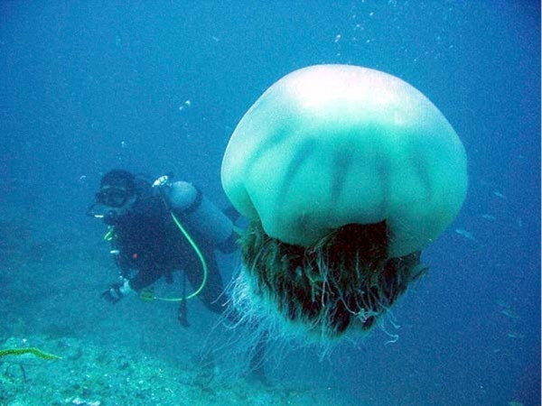
Background: We aimed to study the effects of intra-articular injection of jellyfish mucin (qniumucin) on articular cartilage degeneration in a model of osteoarthritis (OA) created in rabbit knees by resection of the anterior cruciate ligament. Qniumucin was extracted from Aurelia aurita (moon jellyfish) and Stomolophus nomurai (Nomura's jellyfish) and purified by ion exchange chromatography. The OA model used 36 knees in 18 Japanese white rabbits. Purified qniumucin extracts from S. nomurai or A. aurita were used at 1 mg/ml. Rabbits were divided into four groups: a control (C) group injected with saline; a hyaluronic acid (HA)-only group (H group); two qniumucin-only groups (M groups); and two qniumucin + HA groups (MH groups). One milligram of each solution was injected intra-articularly once a week for 5 consecutive weeks, starting from 4 weeks after surgery. Ten weeks after surgery, the articular cartilage was evaluated macroscopically and histologically.
Results: In the C and M groups, macroscopic cartilage defects extended to the subchondral bone medially and laterally. When the H and both MH groups were compared, only minor cartilage degeneration was observed in groups treated with qniumucin in contrast to the group without qniumucin. Histologically, densely safranin-O-stained cartilage layers were observed in the H and two MH groups, but cartilage was strongly maintained in both MH groups.
Conclusion: At the concentrations of qniumucin used in this study, injection together with HA inhibited articular cartilage degeneration in this model of OA.
Figures


Similar articles
-
Quantitative MR T2 measurement of articular cartilage to assess the treatment effect of intra-articular hyaluronic acid injection on experimental osteoarthritis induced by ACLX.Osteoarthritis Cartilage. 2010 Jan;18(1):54-60. doi: 10.1016/j.joca.2009.08.014. Epub 2009 Sep 6. Osteoarthritis Cartilage. 2010. PMID: 19761884
-
Mechanical effects of the intraarticular administration of high molecular weight hyaluronic acid plus phospholipid on synovial joint lubrication and prevention of articular cartilage degeneration in experimental osteoarthritis.Arthritis Rheum. 2003 Jul;48(7):1923-9. doi: 10.1002/art.11172. Arthritis Rheum. 2003. PMID: 12847686
-
In vivo impact on rabbit subchondral bone of viscosupplementation with a hyaluronic acid antioxidant conjugate.BMC Musculoskelet Disord. 2024 Dec 19;25(1):1018. doi: 10.1186/s12891-024-07921-0. BMC Musculoskelet Disord. 2024. PMID: 39702245 Free PMC article.
-
Effect of intraarticular hyaluronan injection on vertical ground reaction force and progression of osteoarthritis after anterior cruciate ligament transection.J Rheumatol. 2005 Feb;32(2):325-34. J Rheumatol. 2005. PMID: 15693095
-
Animal evidence for hyaluronic acid efficacy in knee trauma injuries. Review of animal-model studies.Phys Ther Sport. 2013 May;14(2):116-23. doi: 10.1016/j.ptsp.2013.02.001. Epub 2013 Mar 16. Phys Ther Sport. 2013. PMID: 23506791 Review.
Cited by
-
Novel Marine Organism-Derived Extracellular Vesicles for Control of Anti-Inflammation.Tissue Eng Regen Med. 2021 Feb;18(1):71-79. doi: 10.1007/s13770-020-00319-8. Epub 2021 Jan 7. Tissue Eng Regen Med. 2021. PMID: 33415671 Free PMC article.
-
Evolutionary conservation of the antimicrobial function of mucus: a first defence against infection.NPJ Biofilms Microbiomes. 2018 Jul 4;4:14. doi: 10.1038/s41522-018-0057-2. eCollection 2018. NPJ Biofilms Microbiomes. 2018. PMID: 30002868 Free PMC article. Review.
-
Multigene phylogeny of the scyphozoan jellyfish family Pelagiidae reveals that the common U.S. Atlantic sea nettle comprises two distinct species (Chrysaora quinquecirrha and C. chesapeakei).PeerJ. 2017 Oct 13;5:e3863. doi: 10.7717/peerj.3863. eCollection 2017. PeerJ. 2017. PMID: 29043109 Free PMC article.
-
Jellyfish extract induces apoptotic cell death through the p38 pathway and cell cycle arrest in chronic myelogenous leukemia K562 cells.PeerJ. 2017 Jan 19;5:e2895. doi: 10.7717/peerj.2895. eCollection 2017. PeerJ. 2017. PMID: 28133573 Free PMC article.
-
Bioactive Compounds of Nutraceutical Value from Fishery and Aquaculture Discards.Foods. 2021 Jun 28;10(7):1495. doi: 10.3390/foods10071495. Foods. 2021. PMID: 34203174 Free PMC article. Review.
References
-
- Sato M, Mochida J. Osteoarthritis. Yakkyoku (Journal of Practical Pharmacy) 2007;58:858–865.
-
- Huskisson EC, Berry H, Gishen P, Jubb RW, Whitehead J. Effects of antiinflammatory drugs on the progression of osteoarthritis of the knee. LINK study group. Longitudinal investigation of nonsteroidal antiinflammatory drugs in knee osteoarthritis. J Rheumatol. 1995;22:1941–1946. - PubMed
Publication types
MeSH terms
Substances
LinkOut - more resources
Full Text Sources
