

Current source density (CSD) old/new effects during recognition memory for words and faces in schizophrenia and in healthy adults
- PMID: 19995583
- PMCID: PMC2856653
- DOI: 10.1016/j.ijpsycho.2009.12.001
Current source density (CSD) old/new effects during recognition memory for words and faces in schizophrenia and in healthy adults
Abstract
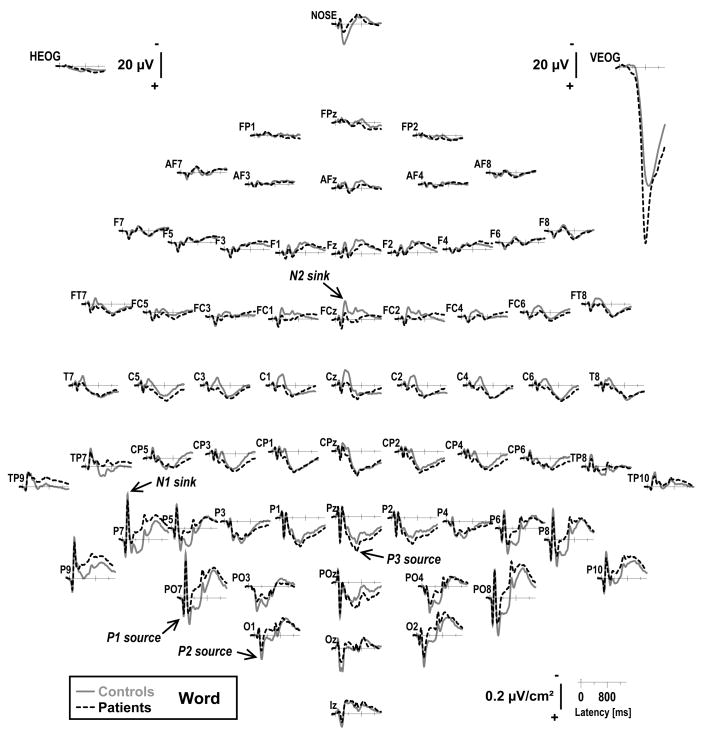
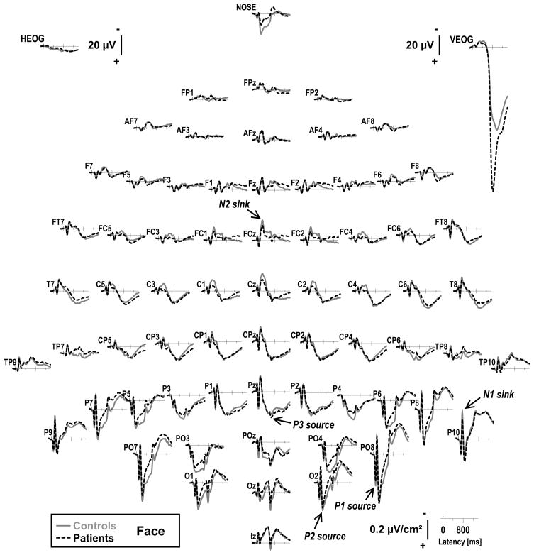
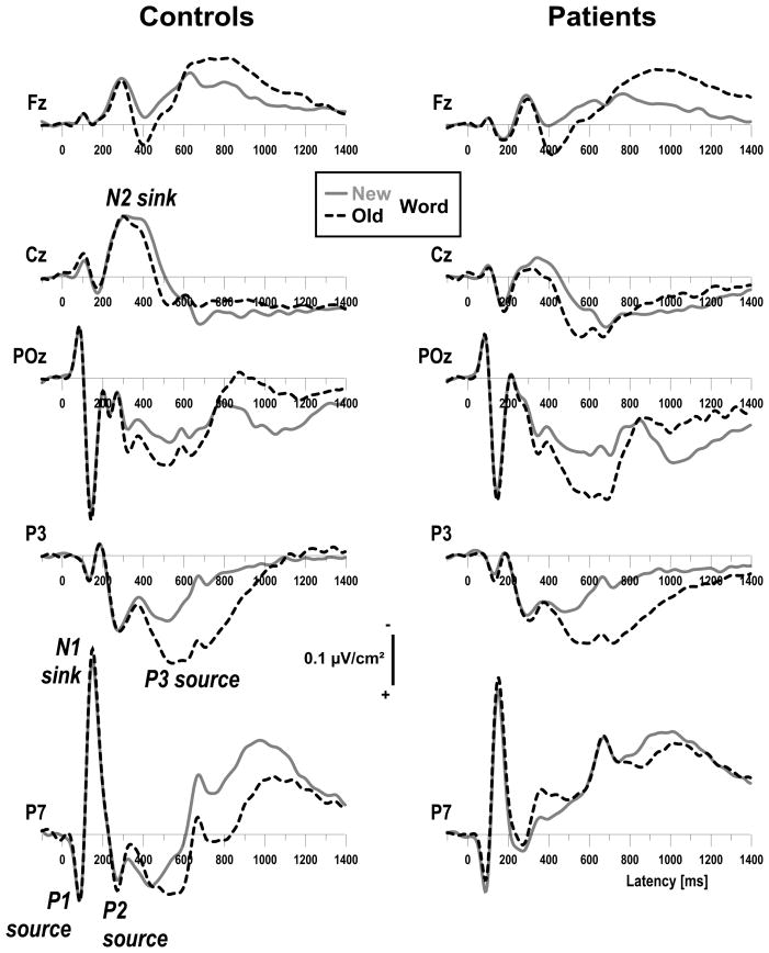
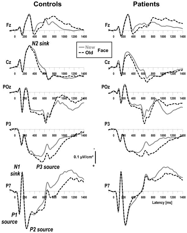
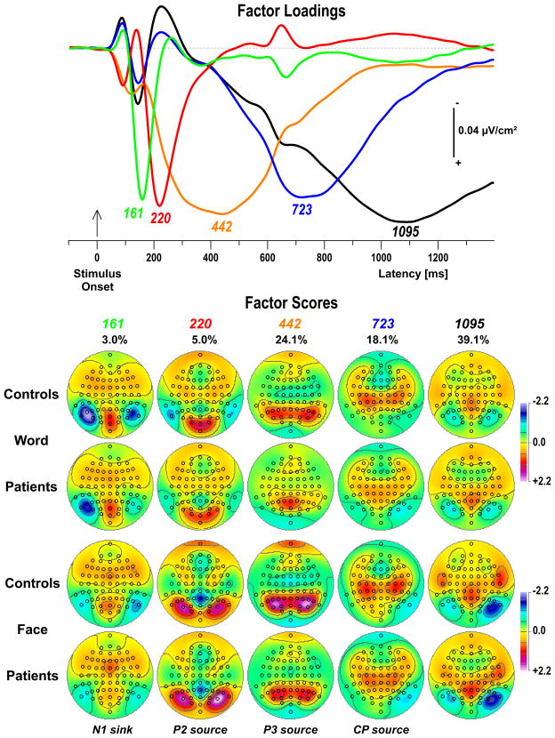
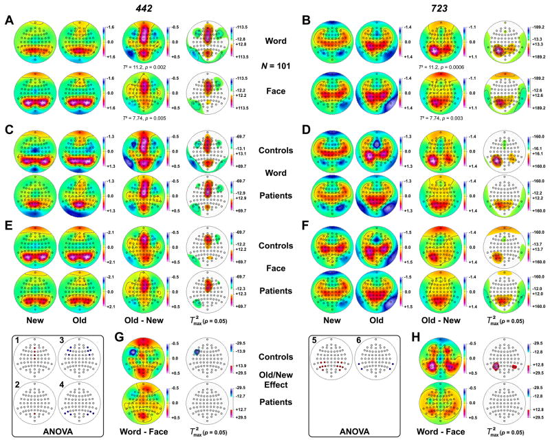
We previously reported a preserved 'old-new effect' (enhanced parietal positivity 300-800 ms following correctly-recognized repeated words) in schizophrenia over mid-parietal sites using 31-channel nose-referenced event-related potentials (ERP) and reference-free current source densities (CSD). However, patients showed poorer word recognition memory and reduced left lateral-parietal P3 sources. The present study investigated whether these abnormalities are specific to words. High-density ERPs (67 channels) were recorded from 57 schizophrenic (24 females) and 44 healthy (26 females) right-handed adults during parallel visual continuous recognition memory tasks using common words or unknown faces. To identify and measure neuronal generator patterns underlying ERPs, unrestricted Varimax-PCA was performed using CSD estimates (spherical spline surface Laplacian). Two late source factors peaking at 442 ms (lateral parietal maximum) and 723 ms (centroparietal maximum) accounted for most of the variance between 250 and 850 ms. Poorer (76.6+/-20.0% vs. 85.7+/-12.4% correct) and slower (824+/-170 vs. 755+/-147 ms) performance in patients was accompanied by reduced stimulus-locked parietal sources. However, both controls and patients showed mid-frontal (442 ms) and left parietal (723 ms) old/new effects in both tasks. Whereas mid-frontal old/new effects were comparable across groups and tasks, later left parietal old/new effects were markedly reduced in patients over lateral temporoparietal but not mid-parietal sites, particularly for words, implicating impaired phonological processing. In agreement with prior results, ERP correlates of recognition memory deficits in schizophrenia suggest functional impairments of lateral posterior cortex (stimulus representation) associated with conscious recollection. This deficit was more pronounced for common words despite a greater difficulty to recall unknown faces, indicating that it is not due to a generalized cognitive deficit in schizophrenia.
Copyright 2009 Elsevier B.V. All rights reserved.
Figures
Similar articles
-
ERP generator patterns in schizophrenia during tonal and phonetic oddball tasks: effects of response hand and silent count.Clin EEG Neurosci. 2010 Oct;41(4):184-95. doi: 10.1177/155005941004100405. Clin EEG Neurosci. 2010. PMID: 21077570 Free PMC article.
-
Stimulus- and response-locked neuronal generator patterns of auditory and visual word recognition memory in schizophrenia.Int J Psychophysiol. 2009 Sep;73(3):186-206. doi: 10.1016/j.ijpsycho.2009.02.003. Epub 2009 Mar 9. Int J Psychophysiol. 2009. PMID: 19275917 Free PMC article.
-
Principal components analysis of Laplacian waveforms as a generic method for identifying ERP generator patterns: I. Evaluation with auditory oddball tasks.Clin Neurophysiol. 2006 Feb;117(2):348-68. doi: 10.1016/j.clinph.2005.08.034. Epub 2005 Dec 13. Clin Neurophysiol. 2006. PMID: 16356767
-
Brain event-related potentials (ERPs) in schizophrenia during a word recognition memory task.Int J Psychophysiol. 1999 Dec;34(3):249-65. doi: 10.1016/s0167-8760(99)00082-3. Int J Psychophysiol. 1999. PMID: 10610049
-
Remembering or knowing: electrophysiological evidence for an episodic memory deficit in schizophrenia.Psychol Med. 2002 Oct;32(7):1261-71. doi: 10.1017/s0033291702006335. Psychol Med. 2002. PMID: 12420895
Cited by
-
A neurophysiological deficit in early visual processing in schizophrenia patients with auditory hallucinations.Psychophysiology. 2012 Sep;49(9):1168-78. doi: 10.1111/j.1469-8986.2012.01404.x. Epub 2012 Jul 16. Psychophysiology. 2012. PMID: 22803512 Free PMC article.
-
ERP generator patterns in schizophrenia during tonal and phonetic oddball tasks: effects of response hand and silent count.Clin EEG Neurosci. 2010 Oct;41(4):184-95. doi: 10.1177/155005941004100405. Clin EEG Neurosci. 2010. PMID: 21077570 Free PMC article.
-
Neuronal generator patterns of olfactory event-related brain potentials in schizophrenia.Psychophysiology. 2010 Nov;47(6):1075-86. doi: 10.1111/j.1469-8986.2010.01013.x. Psychophysiology. 2010. PMID: 20456657 Free PMC article.
-
Smiling as negative feedback affects social decision-making and its neural underpinnings.Cogn Affect Behav Neurosci. 2020 Feb;20(1):160-171. doi: 10.3758/s13415-019-00759-3. Cogn Affect Behav Neurosci. 2020. PMID: 31900873
-
Heterogeneity of auditory verbal working memory in schizophrenia.J Abnorm Psychol. 2011 Feb;120(1):88-97. doi: 10.1037/a0021661. J Abnorm Psychol. 2011. PMID: 21319926 Free PMC article.
References
-
- Alain C, Cortese F, Bernstein LJ, He Y, Zipursky RB. Auditory feature conjunction in patients with schizophrenia. Schizophr Res. 2001;49(1–2):179–191. - PubMed
-
- Alain C, Bernstein LJ, He Y, Cortese F, Zipursky RB. Visual feature conjunction in patients with schizophrenia: an event-related brain potential study. Schizophr Res. 2002;57(1):69–79. - PubMed
-
- Albus M, Hubmann W, Mohr F, Hecht S, Hinterberger Weber P, Seitz NN, Kuchenhoff H. Neurocognitive functioning in patients with first-episode schizophrenia: results of a prospective 5-year follow-up study. Eur Arch Psychiatry Clin Neurosci. 2006;256(7):442–451. - PubMed
-
- Allan K, Wilding EL, Rugg MD. Electrophysiological evidence for dissociable processes contributing to recollection. Acta Psychol (Amst) 1998;98(2–3):231–252. - PubMed
Publication types
MeSH terms
Grants and funding
LinkOut - more resources
Full Text Sources
Medical
