. 2009 Nov;2(6):1384-96.
doi: 10.1093/mp/ssp085. Epub 2009 Oct 30.

Molecular characterization of the Calvin cycle enzyme phosphoribulokinase in the stramenopile alga Vaucheria litorea and the plastid hosting mollusc Elysia chlorotica

Affiliations

Molecular characterization of the Calvin cycle enzyme phosphoribulokinase in the stramenopile alga Vaucheria litorea and the plastid hosting mollusc Elysia chlorotica

Mary E Rumpho et al. Mol Plant. 2009 Nov.

Abstract

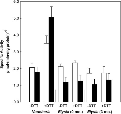
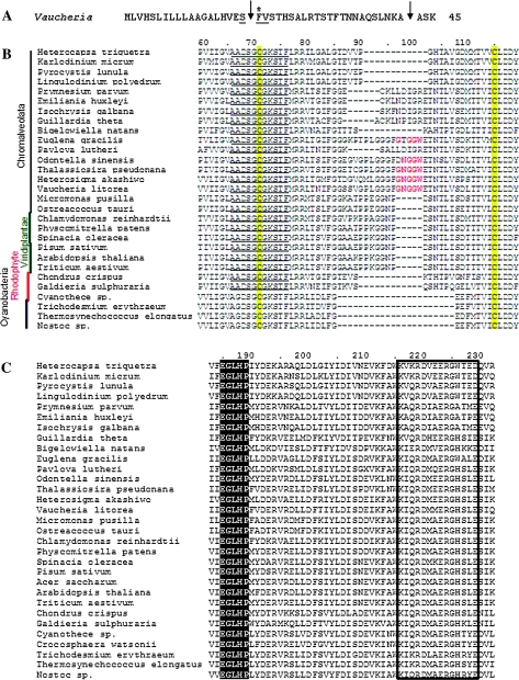
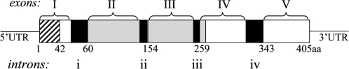
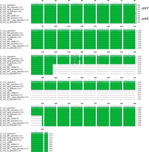
Phosphoribulokinase (PRK), a nuclear-encoded plastid-localized enzyme unique to the photosynthetic carbon reduction (Calvin) cycle, was cloned and characterized from the stramenopile alga Vaucheria litorea. This alga is the source of plastids for the mollusc (sea slug) Elysia chlorotica which enable the animal to survive for months solely by photoautotrophic CO2 fixation. The 1633-bp V. litorea prk gene was cloned and the coding region, found to be interrupted by four introns, encodes a 405-amino acid protein. This protein contains the typical bipartite target sequence expected of nuclear-encoded proteins that are directed to complex (i.e. four membrane-bound) algal plastids. De novo synthesis of PRK and enzyme activity were detected in E. chlorotica in spite of having been starved of V. litorea for several months. Unlike the algal enzyme, PRK in the sea slug did not exhibit redox regulation. Two copies of partial PRK-encoding genes were isolated from both sea slug and aposymbiotic sea slug egg DNA using PCR. Each copy contains the nucleotide region spanning exon 1 and part of exon 2 of V. litorea prk, including the bipartite targeting peptide. However, the larger prk fragment also includes intron 1. The exon and intron sequences of prk in E. chlorotica and V. litorea are nearly identical. These data suggest that PRK is differentially regulated in V. litorea and E. chlorotica and at least a portion of the V. litorea nuclear PRK gene is present in sea slugs that have been starved for several months.

Keywords: Alga; Calvin cycle; Elysia chlorotica; Vaucheria litorea; kleptoplast; mollusc; phosphoribulokinase; photosynthesis; plastid; redox regulation; stramenopile; symbiosis.


Figures

Figure 1.
Figure 1.
Lifecycle Stages of Kleptoplastic Elysia chlorotica. (A) Aposymbiotic sea slug eggs with filaments of Vaucheria litorea in the background. Egg ribbons can vary from 3 to 30 cm in length. (B) Larval stage. Scale bar  =  50 μm. (C) Juvenile kleptoplastic E. chlorotica having fed on V. litorea for 1 d. Scale bar  =  100 μm. (D) Internal structures of adult E. chlorotica showing the close physical contact of the reproductive ovotestes (o), digestive diverticuli (d) containing green kleptoplasts, and the blood vasculature (v). Scale bar  =  250 μm. (E) Dorsal view of a young, adult E. chlorotica revealing the finely divided digestive diverticuli (d) which distributes the kleptoplasts throughout the body, except within the aposymbiotic heart (h). Scale bar  =  500 μm. (F) Mature E. chlorotica illustrating the uniform green coloring throughout the adult body. Scale bar  =  3 mm.
Figure 2.
Figure 2.
Detection of PRK in Elysia chlorotica. (A) PRK Western blot using a tobacco PRK antibody and the alkaline phosphatase detection system. Extracts were loaded on an equal chlorophyll basis as follows: E-kp, E. chlorotica kleptoplasts (from animals starved nine months); V-cp, V. litorea plastids; S-cp, spinach plastids; E-tp, E. chlorotica total proteins (from animals starved for 3 months); V-tp, V. litorea total proteins. (B) Western blot and fluorogram of immunoprecipitated PRK from 5-month-starved sea slugs, following labeling with [35S]methionine/cysteine for 6 h and separation by SDS–PAGE.
Figure 3.
Figure 3.
Redox Activation of PRK in Vaucheria litorea and Elysia chlorotica. Enzyme activity was measured in crude extracts of algal and sea slug samples collected 5 h into the light (white columns) or dark (black columns) period. The sea slugs had been starved for 0 or 3 months, as indicated. The extracts were prepared in the presence (+DTT) or absence (–DTT) of reducing agent and assayed as described in ‘Methods’. The bars indicate the standard error of the mean with n  =  6 for each algal measurement and n  =  3 for each sea slug measurement.
Figure 4.
Figure 4.
Signature Motifs of the PRK Protein (A) N-terminal amino acid sequence of V. litorea PRK pre-protein showing cleavage sites for predicted signal sequence (first arrow; amino acids 1–19) and target peptide sequence (second arrow, amino acids 20–42). The first site is identified by the conserved motif (SFV) at the border of the ER signal sequence (underlined) and a critical Phe (starred). (B) Clustal W alignment of partial PRK amino acid sequences from different organisms aligned using default parameters. The highly conserved nucleotide binding motif A is shown in bold underline and the two conserved regulatory Cys residues are highlighted in yellow. A 5-amino acid insertion between the Cys residues is shown in red for the Vaucheria sequence and four other chromalveolate sequences. (C) Clustal W alignment of partial PRK amino acid sequences illustrating the highly conserved Walker B motif (black highlighted) and the PRK signature sequence (boxed). Numbers along the sequences indicate the respective amino acid positions relative to the V. litorea sequence beginning with the start Met.
Figure 5.
Figure 5.
Schematic Representation of Functional Features of the V. litorea PRK Protein Imposed Upon the Corresponding Gene Organization. Five exons enclosed in brackets (I–V) and four introns represented by black boxes (i–iv) illustrate the genomic structure. Numbered amino acid residues are shown below the corresponding genomic segments. The hatched box represents the plastid transit peptide sequence within exon 1, corresponding to amino acids 1–42. The gray shaded boxes represent the PRK domain sequence, corresponding to amino acids 62–266, and spanning much of exon II, all of exon III, and 24 bp of exon IV.
Figure 6.
Figure 6.
PCR Amplification of prk from V. litorea and E. chlorotica. (A) PCR amplification of prk from algal and sea slug DNA using primers PRK5F and PRK5R. Lane 1, 1 kb Plus DNA ladder (Invitrogen); lane 2, no DNA (negative control); lane 3, pVA1prk construct (pGEM-T Easy vector with the V. litorea prk cDNA as insert; positive control); lane 4, pVA2prk construct (pGEM-T Easy vector with genomic V. litorea prk as insert); and lane 5, E. chlorotica DNA as template. (B) PCR amplification of the prk gene from sea slug egg DNA using primers PRK5F and PRK5R. Lane 1, 1 kb plus ladder; lane 2, no DNA (negative control); lane 3, sea slug egg DNA. The arrows in both figures point to prkY (421 bp) and prkX (269 bp) fragments.
Figure 7.
Figure 7.
Alignment of Partial prk Sequences from V. litorea and E. chlorotica. Clustal W alignment of nucleotide sequences amplified from V. litorea cDNA and genomic DNA, E. chlorotica total DNA from Martha's Vineyard (MV) and Halifax (H) collections, E. chlorotica (MV) egg DNA, and E. chlorotica (MV) cDNA using PRK5F and PRK5R primers (see corresponding PCR bands in Figure 6A and 6B). Numbers in parentheses after the organism name refer to the number of sequences analyzed for each. Numbers at the end of each line indicate the respective nucleotide positions of the amplified product beginning with the start codon ATG. A dot indicates that the residues in that column are identical in all sequences in the alignment. A wavy dash (∼) marks the intron region. W  =  A or T. The larger, intron-containing sequence was labeled prkY, whereas the shorter sequence was labeled prkX.
Figure 8.
Figure 8.
Maximum Likelihood (PhyML) Phylogenetic Analysis of PRK in Cyanobacteria, Algae, and Plants. The results of bootstrap analyses are shown with the PhyML bootstrap values above and the RAxML bootstrap values below the branches. The branch lengths in the trees are proportional to the number of substitutions per site (see scale in figure). The branch uniting the cyanobacteria was used to root this phylogeny.

Similar articles

Cited by

References

    1. Armbrust EV, et al. The genome of the diatom Thalassiosira pseudonana: ecology, evolution, and metabolism. Science. 2004;306:79–86. - PubMed
    1. Bhattacharya D, Yoon HS, Hackett JD. Photosynthetic eukaryotes unite: endosymbiosis connects the dots. Bioessays. 2004;26:50–60. - PubMed
    1. Bock R, Timmis JN. Reconstructing evolution: gene transfer from plastids to the nucleus. Bioessays. 2008;30:556–566. - PubMed
    1. Boggetto N, Gontero B, Maberly SC. Regulation of phosphoribulokinase and glyceraldehyde 3-phosphate dehydrogenase in a freshwater diatom, Asterionella formosa. J. Phycol. 2007;43:1227–1235. - PubMed
    1. Chen XF, et al. Molecular cloning and expression analysis of rice phosphoribulokinase gene that is regulated by environmental stresses. Mol. Biol. Rep. 2004;31:249–255. - PubMed

Publication types

MeSH terms

Substances

Associated data