Review
. 2010 Feb 15;588(Pt 4):565-72.
doi: 10.1113/jphysiol.2009.183160. Epub 2009 Dec 7.

Atomic structure and dynamics of pentameric ligand-gated ion channels: new insight from bacterial homologues

Affiliations
Review

Atomic structure and dynamics of pentameric ligand-gated ion channels: new insight from bacterial homologues

Pierre-Jean Corringer et al. J Physiol. .

Abstract

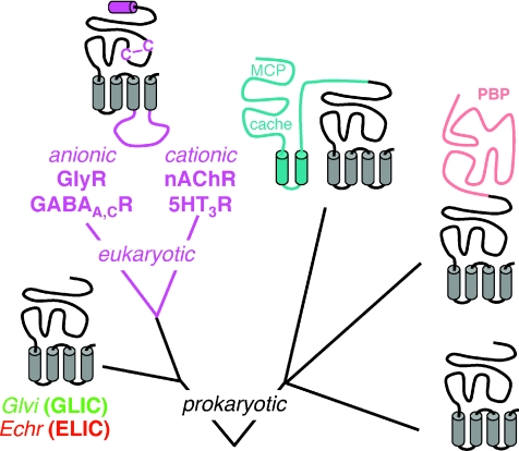
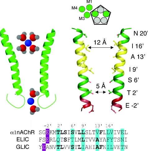
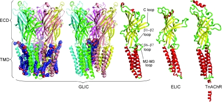
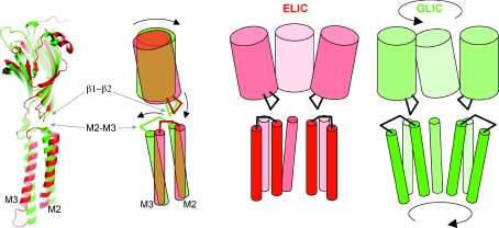
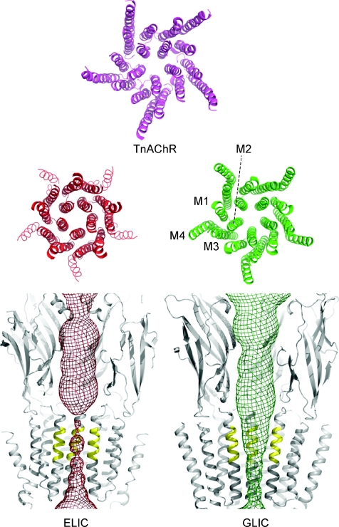
Pentameric ligand-gated ion channels (pLGICs) are widely expressed in the animal kingdom and are key players of neurotransmission by acetylcholine (ACh), gamma-amminobutyric acid (GABA), glycine and serotonin. It is now established that this family has a prokaryotic origin, since more than 20 homologues have been discovered in bacteria. In particular, the GLIC homologue displays a ligand-gated ion channel function and is activated by protons. The prokaryotic origin of these membrane proteins facilitated the X-ray structural resolution of the first members of this family. ELIC was solved at 3.3 A in a closed-pore conformation, and GLIC at up to 2.9 A in an apparently open-pore conformation. These data reveal many structural features, notably the architecture of the pore, including its gate and its selectivity filter, and the interactions between the protein and lipids. In addition, comparison of the structures of GLIC and ELIC hints at a mechanism of channel opening, which consists of both a quaternary twist and a tertiary deformation. This mechanism couples opening-closing motions of the channel with a global reorganization of the protein, including the subunit interface that holds the neurotransmitter binding sites in eukaryotic pLGICs.


Figures

None
Pierre-Jean Corringer trained as a chemist and did his PhD (Paris) and post-doctoral fellowship (Brighton) in organic synthesis. He then joined the Pasteur Institute as a CNRS researcher to work on the functional architecture and biosynthesis of nicotinic acetylcholine receptors. His work contributed to the discovery of bacterial homologues of these neurotransmitter receptors. In 2008 he created his own research group on Channel Receptors in the Pasteur Institute, which has already produced, in collaboration with Marc Delarue, one of the first atomic resolution structures of the bacterial homologues of the nicotinic receptors.
Figure 1
Figure 1. Schematic phylogenetic tree of the pLGIC family
The different subfamilies of genes are shown as cartoons representing their transmembrane topology (transmembrane helices as vertical cylinders). The minimal structure required for ligand-gated ion channel function is shown in grey, and additional modules are shown in colour.
Figure 3
Figure 3. The ion channel of GLIC
Right: cartoon representation of the ion channel of GLIC. Only two M2 segments are represented for clarity, with rings of charged residues in red, of polar residues in green and of hydrophobic residues in yellow. Left: hypothesized ion selection mechanism proposed on the basis of mutational analysis; adapted from Corringer et al. 1999.
Figures 2
Figures 2. X-ray structure of GLIC and comparison with ELIC and nAChR
Left, cartoon representation of the X-ray structure of the GLIC protein at 2.9 Å resolution. Each subunit is coloured differently, and lipids partially seen in the electron density map are represented as spheres. The panel on the right shows the same structure with the bundle of detergent (dodecyl-β-d-maltopyranoside) bound within the ion channel. Right: cartoon representation of a single subunit of GLIC, ELIC and nAChR (TnAChR, from EM data of the Torpedo receptor). Subunits are coloured according to the secondary structure, illustrating the common core of β-sandwich (yellow) and α-helices (red) that is conserved throughout the family, while connecting loops (green) are on the whole highly divergent in structure.
Figure 5
Figure 5. A twist/deformation mechanism for channel opening
Left, superimposition of the GLIC (green) and ELIC (red) structures. Only one subunit is shown, and the tertiary deformation is schematically represented on the right. From ELIC to GLIC, the extracellular β-sandwich undergoes an 8 deg rotation, which, apparently through the interaction between the β1–β2 and M2–M3 loop, is concomitant with a rigid-bogy tilt of the M2 and M3 helices, thereby opening the pore. Right, scheme showing the transition of three subunits together, illustrating the twist motion, with opposite rotations in the upper and lower parts of the protein.
Figure 4
Figure 4. The permeation pathway of GLIC and ELIC
Mesh representation of the aqueous pathway in the ELIC and GLIC structures, illustrating that ELIC is in a closed conformation, and GLIC is apparently open. The top view shows the transmembrane helices alone, illustrating that the conformation of the pore and of the M2 helices are similar between GLIC and TnAChR.

Similar articles

Cited by

References

    1. Bali M, Akabas MH. The location of a closed channel gate in the GABAA receptor channel. J Gen Physiol. 2007;129:145–159. - PMC - PubMed
    1. Beckstein O, Sansom MSP. A hydrophobic gate in an ion channel: the closed state of the nicotinic acetylcholine receptor. Phys Biol. 2006;3:147–159. - PubMed
    1. Bouzat C, Gumilar F, Spitzmaul G, Wang H, Rayes D, Hansen SB, Taylor P, Sine SM. Coupling of agonist binding to channel gating in an ACh-binding protein linked to an ion channel. Nature. 2004;430:896–900. - PubMed
    1. Bocquet N, Prado de Carvalho L, Cartaud J, Neyton J, Poupon CL, Taly A, Grutter T, Changeux J, Corringer PJ. A prokaryotic proton-gated ion channel from the nicotinic acetylcholine receptor family. Nature. 2007;445:116–119. - PubMed
    1. Bocquet N, Nury H, Baaden M, Poupon CL, Changeux J, Delarue M, Corringer PJ. X-ray structure of a pentameric ligand-gated ion channel in an apparently open conformation. Nature. 2009;457:111–114. - PubMed

Publication types

MeSH terms

LinkOut - more resources