. 2010 Jan 15;184(2):764-74.
doi: 10.4049/jimmunol.0903006. Epub 2009 Dec 7.

STAT3 signaling in CD4+ T cells is critical for the pathogenesis of chronic sclerodermatous graft-versus-host disease in a murine model

Affiliations

STAT3 signaling in CD4+ T cells is critical for the pathogenesis of chronic sclerodermatous graft-versus-host disease in a murine model

Vedran Radojcic et al. J Immunol. .

Abstract

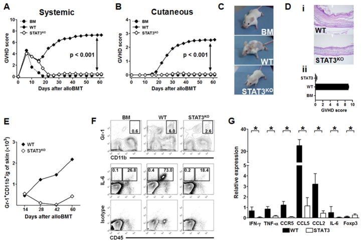
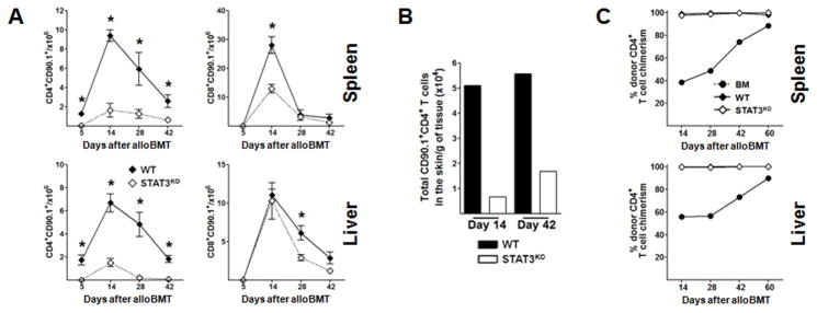
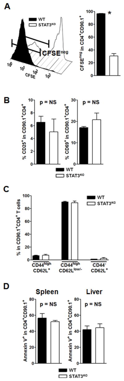
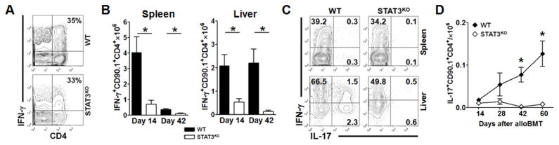
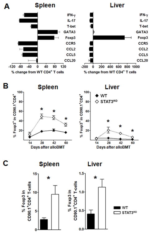
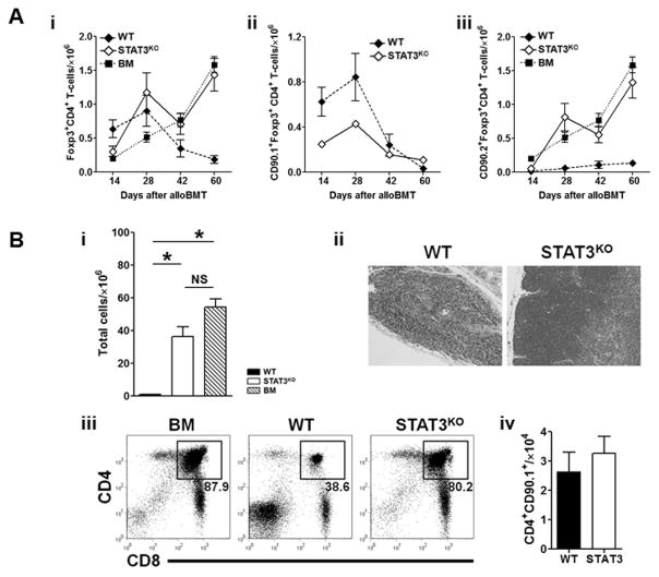
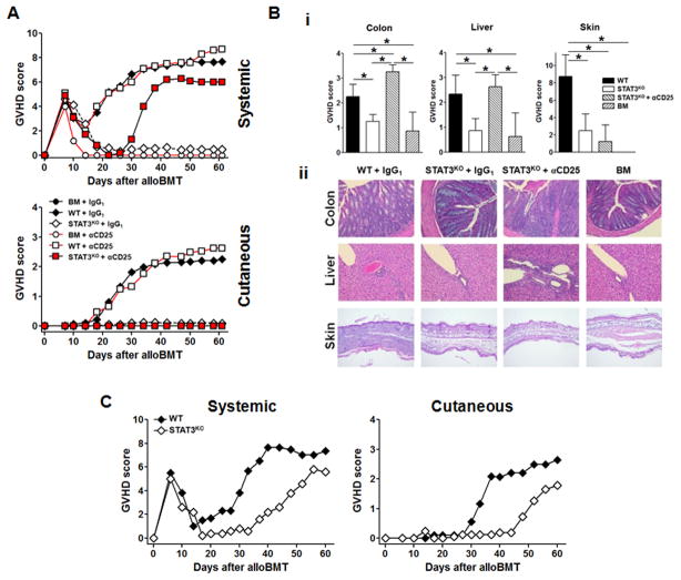
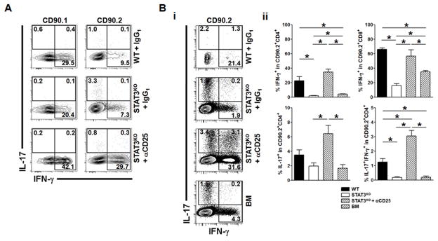
Donor CD4+ T cells are thought to be essential for inducing delayed host tissue injury in chronic graft-versus-host disease (GVHD). However, the relative contributions of distinct effector CD4+ T cell subpopulations and the molecular pathways influencing their generation are not known. We investigated the role of the STAT3 pathway in a murine model of chronic sclerodermatous GVHD. This pathway integrates multiple signaling events during the differentiation of naive CD4+ T cells and impacts their homeostasis. We report that chimeras receiving an allograft containing STAT3-ablated donor CD4+ T cells do not develop classic clinical and pathological manifestations of alloimmune tissue injury. Analysis of chimeras showed that abrogation of STAT3 signaling reduced the in vivo expansion of donor-derived CD4+ T cells and their accumulation in GVHD target tissues without abolishing antihost alloreactivity. STAT3 ablation did not significantly affect Th1 differentiation while enhancing CD4+CD25+Foxp3+ T cell reconstitution through thymus-dependent and -independent pathways. Transient depletion of CD25+ T cells in chimeras receiving STAT3-deficient T cells resulted in delayed development of alloimmune gut and liver injury. This delayed de novo GVHD was associated with the emergence of donor hematopoietic stem cell-derived Th1 and Th17 cells. These results suggest that STAT3 signaling in graft CD4+ T cells links the alloimmune tissue injury of donor graft T cells and the emergence of donor hematopoietic stem cell-derived pathogenic effector cells and that both populations contribute, albeit in different ways, to the genesis of chronic GVHD after allogenic bone marrow transplantation in a murine model.


Figures

Figure 1
Figure 1. STAT3 signaling in CD4+ T-cells is critical for the development of chronic GVHD in B10.D2→BALB/c model
On day 0, cohorts of BALB/c-CD45.1 recipients were lethally irradiated and reconstituted with T cell depleted (TCD) CD90.2+ BM only (●), TCD BM with 1.8 × 10⁶ WT CD90.1+CD4+ and 0.9 × 10⁶ WT CD90.1+CD8+ T-cells (WT GVH inoculum, ◆), or 1.8 × 10⁶ STAT3KO CD90.1+CD4+ and 0.9 × 10⁶ WT CD90.1+CD8+ T-cells (STAT3KO GVH inoculum, ◇) from B10.D2 donors. All mice were monitored for clinical signs of GVHD using systemic or skin-specific GVHD scoring systems. (A) Mean systemic clinical GVHD scores. p < 0.001 (◆vs. ◇, and ◆ vs. ●); p = NS (● vs. ◇). (B) Mean skin-specific clinical GVHD scores; p < 0.001 (◆ vs. ◇, and ◆ vs. ●); p = NS (● vs. ◇). Experiment was repeated at least six times, with total of more than 50 animals per cohort. (C) Representative photographs taken on day 60 after alloBMT show surviving chimeras that received TCD BM only, WT, or STAT3KO GVH inocula. (D) i-Representative photomicrographs of histopathological changes in the skin of chimeras depicted in panel C (hematoxylin and eosin stain, original magnification ×200). Note the presence of diffuse epidermal thickening and increased dermal cellularity due to lymphoid infiltration in the skin of WT CD4+ recipients compared to normal-appearing skin of STAT3KO GVH inoculum recipients. ii-GVHD skin scores on day 60 after alloBMT; n=5 chimeras per group (E–F) Skin was harvested from chimeras and single cell suspensions were prepared for flow cytometric analysis of infiltrating inflammatory Gr-1+CD11b+ monocytes (E–F) and production of IL-6 (F). (WT–◆, STAT3KO – ◇). (G) Gene expression levels of proinflammatory cytokines, chemokines and their receptors and transcription factors in the skin were determined by real-time quantitative RT-PCR. Obtained values were normalized to 18s rRNA expression. * indicates p < 0.05. Data are expressed as mean ± SD.
Figure 2
Figure 2. STAT3 ablation abrogates GVHD without impairing achievement of complete donor chimerism
Lethally irradiated BALB/c-CD45.1 recipients were reconstituted with TCD BM plus WT or STAT3KO GVH inocula. To monitor the fate of graft-derived T cells we utilized TCD BM from B10.D2-Thy1.2 mice while all T cells were from donors on a B10.D2-Thy1.1 background. (A) Serial analysis of absolute numbers of graft-derived CD90.1+ T-cells in spleens and livers of chimeras that received WT or STAT3KO inocula. Data are representative of four independent experiments with a total of more than 12 animals per group and time-point. Results are presented as mean ± SEM; * indicates p < 0.05. (B) Analysis of CD90.1+CD4+ T-cells in the skin on days 14 and 42 after alloBMT. (C) Changes in donor chimerism were serially monitored in spleen and liver of chimeras that received TCD BM (●), or TCD BM plus WT (◆) or STAT3KO (◇) GVH inocula based on the differential expression of CD45 on donor B10.D2 (CD45.2+) and host (CD45.1+) T-cells. Note no difference in the chimerism levels between WT and STAT3KO chimeras at all time-points evaluated.
Figure 3
Figure 3. STAT3 ablation in CD4+ T-cells influences their in vivo proliferation and expansion
BALB/c-CD45.1 recipients were lethally irradiated and reconstituted with WT or STAT3KO GVH inocula containing CFSE-labeled graft-derived CD90.1+ T-cells. (A) Representative histogram plot and CFSE dilution profile of graft-derived spleen-infiltrating CD4+ T-cells on day 5 after adoptive transfer. (* indicates p < 0.01). (B–C) Expression of CD25/CD69 (B), and CD44/CD62L (C) on graft-derived CFSElow CD4+ T-cells retrieved from WT and STAT3KO chimeras on day 14 after alloBMT. (p = NS, for all valid comparisons). (D) In vivo analysis of apoptosis of WT and STAT3KO CD4+ T-cells. On day 5 after alloBMT splenic and hepatic graft-derived CD4+ T-cells were harvested and stained for Annexin V. Representative data from one out of three independent experiments are shown.
Figure 4
Figure 4. Abrogation of STAT3 signaling prevents late emergence of Th17 cells while minimally affecting Th1 response
Lethally irradiated BALB/c-CD45.1 recipients were reconstituted with TCD BM, or TCD BM with WT or STAT3KO GVH inocula. Skin, splenic and liver mononuclear cells were serially retrieved and analyzed for cytokine production by flow cytometry. (A) Percentage of IFN-γ producing graft-derived CD90.1+CD4+ T-cells in skin of chimeras (day 14 after alloBMT). (B) Production of IFN-γ in graft derived CD4+ T-cells on days 14 and 42 after alloBMT. (C) Representative contour plots showing IFN-γ and IL-17 production in graft-derived CD4+ T-cells harvested from spleens and livers of WT and STAT3KO chimeras 42 days after alloBMT. (D) Serial analysis of IL-17 production in graft derived CD4+ T-cells in the liver points to their emergence later during GVHD evolution. * indicates p < 0.05 for WT vs. STAT3KO GVH inoculum recipients. Data are representative of at least three experiments with >15 mice per group.
Figure 5
Figure 5. Targeted ablation of STAT3 signaling in donor CD4+ T-cells augments expansion of Foxp3+CD4+ Tregs
Lethally irradiated BALB/c-CD45.1 mice received TCD BM alone or TCD BM plus WT or STAT3KO GVH inocula. (A) Gene expression analysis of sorted graft-derived (CD90.1+) CD4+ T-cells from spleens and livers of chimeras on day 42 after alloBMT. Data are presented as percent change from the expression level of a given gene in WT CD4+ T-cells using the formula: %change=100×(STAT3KOrelativeexpression)/(WTrelativeexpression¯), where relative expression = 2[Ct targetCt housekeeping gene]. (B) Longitudinal analysis of graft-derived CD90.1+CD4+ T-cells in spleen and liver shows increased polarization of STAT3KO CD4+ T-cells toward the CD4+Foxp3+ Treg phenotype. * indicates p < 0.05 for WT vs. STAT3KO GVH inoculum recipients. (C) Increased conversion of STAT3KO CD90.1+ CD4+CD25 T-cells. STAT3KO and WT CD4+CD25- were purified by FACS and combined with donor CD8+ T-cells and TCD BM and given as a part of a GVH inoculum. On day 28 after alloBMT spleen and livers of chimeras were harvested and percentage of CD90.1+CD4+CD25+Foxp3+ T cells was determined by flow cytometry. * indicates p < 0.05 for WT vs. STAT3KO GVH inoculum recipients. Data from two independent experiments with >9 analyzed mice per group are shown
Figure 6
Figure 6. Unperturbed reconstitution of donor HSC-derived Tregs is a dominant feature in chimeras receiving STAT3KO GVH inocula
Irradiated BALB/c-CD45.1 recipients were transplanted with TCD BM, or TCD BM plus WT or STAT3KO GVH inocula. (A) Longitudinal analysis of total splenic (i), CD90.1+ graft- (ii), and CD90.2+ HSC- (iii) derived Foxp3+CD4+ Treg reconstitution. (B) Thymic damage is uniquely present in WT GVH inoculum recipients. i-Thymic cellularity (* indicates p < 0.05; WT vs. STAT3). ii-Histopathological analysis (hematoxylin and eosin stain, original magnification ×200). Note destruction of thymic epithelium, lack of delineation between cortical and medullary areas and severe atrophy in thymuses harvested from chimeras that receiving WT GVH inocula. iii-Representative dot plots. Numbers within the gates indicate mean percentage of double positive donor-derived CD4+/CD8+ thymocytes. iv-Thymic infiltration by CD90.1+CD4+ T-cells. All data are representative of at least two independent experiments with more than 10 chimeras per group analyzed.
Figure 7
Figure 7. In vivo CD25 depletion or thymectomy induces GVHD in recipients of STAT3KO GVH inocula
Cohorts of BALB/c recipients were irradiated and reconstituted with TCD BM, or TCD BM plus WT or STAT3KO GVH inocula. Designated groups received 0.5 mg of anti-CD25 (TCD BM only– ○; WT – □; STAT3KO–red squares) or isotype control (IgG1; TCD BM only – ●; WT – ◆; STAT3KO –◇) Ab intraperitoneally every third day for a total of 6 injections, beginning on day 1 after alloBMT. (A) Mean systemic and cutaneous GVHD clinical scores. Cumulative data from three independent experiments with >15 animals per group are shown.; p < 0.001 for both groups of □ and ◆ vs. ◇, ○, and ● from day 14 onwards; p < 0.05 for red squares vs. ◇, ○, and ● from day 34 onwards; p = NS for □ vs. ◆ for all time-points analyzed, and for ◆ vs. red squares from day 38 onwards. (B) Histology of colon, liver and skin. i-Overall pathological scores for a given organ. Tissues were collected for histopathological analysis on day 28 after alloBMT just prior to occurrence of clinical signs of GVHD. Data are presented as means ± SD; * indicates p < 0.05. ii-Representative photomicrographs. Note extensive intermucosal infiltration of the lamina propria with decreased goblet cells in the colon, as well as perivascular and peribiliary duct inflammation in the liver in chimeras receiving WT GVH inocula with IgG1, and as well as STAT3KO GVH inocula with anti-CD25. Epidermal hyperplasia and dermal and subdermal inflammation in the skin with destruction of the fatty layer were present only in mice receiving WT CD4+ T-cells. Sections stained with hematoxylin and eosin, original magnification ×200. (C) STAT3KO CD4+ T-cells are able to induce all signs of chronic sclerodermatous GVHD in the absence of the host’s thymus. Thymectomized BALB/c animals were transplanted with STAT3KO or WT GVH inocula (n=6 per group) and serially monitored for signs of systemic and cutaneous GVHD. Note the delayed occurrence of all signs of GVHD in STAT3KO GVH inoculum recipients. For systemic GVHD, p < 0.05 for ◆ vs. ◇ from day 20 onward; for cutaneous GVHD, p < 0.05 for ◆ vs. ◇ for days 34 to 54.
Figure 8
Figure 8. Both HSC-derived Th1 and Th17 are mediators of GVHD in chimeras treated with PC61 mAb
Groups of chimeras were constructed and treated with anti-CD25 or isotype control Abs as described in Figure 7. On day 28 after alloBMT, spleen and mesenteric lymph nodes were retrieved and analyzed for production of IFN-γ and IL-17 in graft (CD90.1+)- and HSC (CD90.2+)-derived T-cells. (A) Representative contour plots show the percentage of IFN-γ and IL-17-secreting cells within the gated graft- and HSC-derived CD4+ T-cells harvested from spleens of designated chimeras. Note the difference in IFN-γ production between anti-CD25 and isotype-treated STAT3KO GVH inoculum recipients. (B) Production of IFN-γ and/or IL-17 in donor HSC-derived CD4+ T-cells harvested from MLNs. i-Representative contour plots depicting cytokine secretion profile of CD90.2+ CD4+ T-cells. Profound effects of CD25 depletion are most evident in the emergence of both Th1 and Th17 HSC-derived CD4+ T-cells. ii-Mean percentage ± SEM of IFN-γ and/or IL-17-secreting CD4+ and CD8+ CD90.2+ HSC-derived T cells; * indicates p < 0.05. Cumulative results of two independent experiments with a total of >10 animals/group analyzed are presented.

Similar articles

Cited by

References

    1. Ferrara JL, Reddy P. Pathophysiology of graft-versus-host disease. Semin Hematol. 2006;43:3–10. - PubMed
    1. Shlomchik WD. Graft-versus-host disease. Nat Rev Immunol. 2007;7:340–352. - PubMed
    1. Parkman R. Is chronic graft versus host disease an autoimmune disease? Curr Opin Immunol. 1993;5:800–803. - PubMed
    1. Tivol E, Komorowski R, Drobyski WR. Emergent autoimmunity in graft-versus-host disease. Blood. 2005;105:4885–4891. - PMC - PubMed
    1. Zhang Y, Hexner E, Frank D, Emerson SG. CD4+ T Cells Generated De Novo from Donor Hemopoietic Stem Cells Mediate the Evolution from Acute to Chronic Graft-versus-Host Disease. J Immunol. 2007;179:3305–3314. - PubMed

Publication types

MeSH terms

Substances