. 2009 Dec 14;187(6):773-80.
doi: 10.1083/jcb.200903033.

Overall Cdk activity modulates the DNA damage response in mammalian cells

Affiliations

Overall Cdk activity modulates the DNA damage response in mammalian cells

Antonio Cerqueira et al. J Cell Biol. .

Abstract

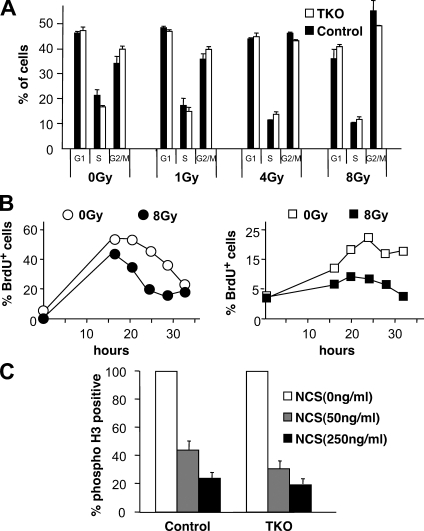
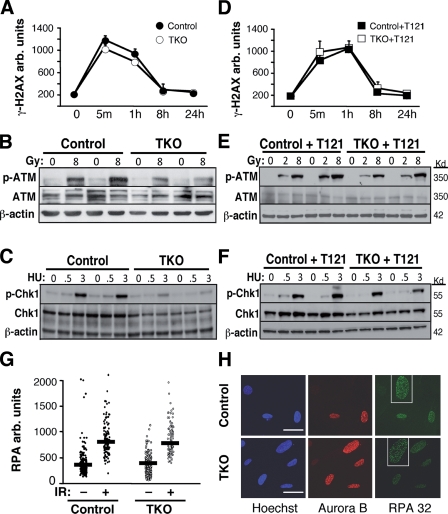
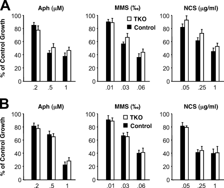
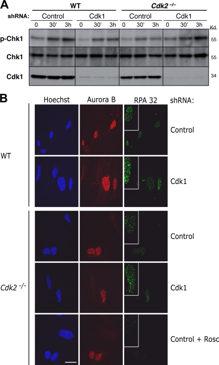
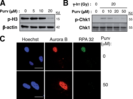
In response to DNA damage, cells activate a phosphorylation-based signaling cascade known as the DNA damage response (DDR). One of the main outcomes of DDR activation is inhibition of cyclin-dependent kinase (Cdk) activity to restrain cell cycle progression until lesions are healed. Recent studies have revealed a reverse connection by which Cdk activity modulates processing of DNA break ends and DDR activation. However, the specific contribution of individual Cdks to this process remains poorly understood. To address this issue, we have examined the DDR in murine cells carrying a defined set of Cdks. Our results reveal that genome maintenance programs of postreplicative cells, including DDR, are regulated by the overall level of Cdk activity and not by specific Cdks.


Figures

Figure 1.
Figure 1.
Functional G1/S and G2/M checkpoints in the absence of interphase Cdks. (A) Cell cycle distribution of Cdk4+/+;Cdk2+/+;Cdk6−/− control and TKO MEFs 10 h after IR with the indicated doses. (B) Percentage of quiescent Cdk4+/+;Cdk2+/+;Cdk6−/− control (left) and TKO (right) MEFs in S phase after serum stimulation and 2-h pulses of BrdU. Cells were either nonirradiated or exposed to 8 Gy of IR before serum stimulation. (C) Fraction of phospho-H3–positive Cdk4+/+;Cdk2+/+;Cdk6−/− control and TKO MEFs either untreated or 45 min after NCS. Error bars indicate mean ± SD (n = 3).
Figure 2.
Figure 2.
DNA repair and checkpoint activation in TKO MEFs. (A) Cells were treated for 1 h with 50 ng/ml NCS, washed, and analyzed by high throughput microscopy over time. The intensity of the γ-H2AX signal per nucleus was measured for Cdk4+/+;Cdk2+/+;Cdk6−/− control and TKO MEFs at the indicated times. (B) Whole cell extracts were prepared from Cdk4+/+;Cdk2+/+;Cdk6−/− control and TKO MEFs 45 min after 8 Gy of IR. Extracts were blotted with antibodies against ATM-Ser1981 or ATM as indicated in Materials and methods. Samples from two independent experiments were loaded. (C) Cdk4+/+;Cdk2+/+;Cdk6−/− control and TKO MEFs were treated for 2 h with hydroxyurea (HU) at the indicated concentrations (millimolars). Extracts were blotted with antibodies against Chk1-Ser345 or Chk1 as indicated in Materials and methods. Samples from two independent experiments were loaded. (D) Cdk4+/+;Cdk2+/+;Cdk6−/− control and TKO MEFs were infected with retroviral particles expressing the T121 fragment (remaining information is as described in A). (E) Whole cell extracts were prepared from Cdk4+/+;Cdk2+/+;Cdk6−/− control and TKO MEFs expressing the T121 fragment 45 min after IR with 2 and 8 Gy. Extracts were blotted with antibodies against ATM-Ser1981 or ATM as indicated in Materials and methods. Samples from two independent experiments were loaded. (F) Cdk4+/+;Cdk2+/+;Cdk6−/− control and TKO MEFs expressing the T121 fragment were treated for 2 h with hydroxyurea at the indicated concentrations (millimolars). Extracts were blotted with antibodies against Chk1-Ser345 or Chk1 as indicated in Materials and methods. Samples from two independent experiments were loaded. (G) Cdk4+/+;Cdk2+/+;Cdk6−/− control and TKO MEFs were preextracted 3 h after IR, and chromatin-bound RPA were analyzed by high throughput microscopy. The results are the mean of three independent experiments. Horizontal bars mark median values. (H) Cdk4+/+;Cdk2+/+;Cdk6−/− control and TKO MEFs were submitted to IR, maintained in culture for 3 h, and preextracted before incubation with the indicated antibodies and confocal analysis. Insets on RPA32 fields show magnified views of a positive cell. Error bars indicate mean ± SD (n = 3). Bars, 15 µM.
Figure 3.
Figure 3.
TKO MEFs are not hypersensitive to DNA-damaging agents. (A) Cdk4+/+;Cdk2+/+;Cdk6−/− control and TKO MEFs were grown for 5 d in the presence of aphidicolin (Aph), MMS, or NCS at the indicated concentrations. Graphs represent the variation in cell number at the end of the experiment normalized to the untreated controls. The results are the mean of two (control MEFs) and four (TKO MEFs) independent experiments. (B) Cdk4+/+;Cdk2+/+;Cdk6−/− control and TKO MEFs expressing the T121 fragment (remaining information is as described in A). Error bars indicate ± SD.
Figure 4.
Figure 4.
Cdk1 and Cdk2 are dispensable for the onset of the DDR. (A) Wild-type (WT) and Cdk2−/− MEFs were infected with lentiviral vectors expressing a scramble control or an shRNA against Cdk1. Extracts were prepared at the indicated time points after IR and blotted with antibodies against Chk1-Ser345 or Chk1 as indicated in Materials and methods. Cdk1 is shown as depletion control. Black lines indicate that intervening lanes have been spliced out. (B) MEFs were infected with the indicated shRNAs, subjected to IR, maintained in culture for 3 h, and preextracted before incubation with the indicated antibodies and confocal analysis. Insets on RPA32 fields show magnified views of a positive cell. (bottom) Cdk2−/− MEFs were preincubated for 3 h before IR with 50 µM of the Cdk inhibitor roscovitine (Rosc). Bar, 15 µM.
Figure 5.
Figure 5.
The Cdk inhibitor purvalanol abrogates DSB end processing. (A) TKO MEFs were treated for 2 h with the indicated concentrations of the Cdk inhibitor purvalanol (Purv). Extracts were blotted with a phosphospecific antibody against histone H3–Ser10. (B) Purvalanol-treated cells were subjected to IR, and extracts were prepared after 30 min. Extracts were blotted with antibodies against Chk1-Ser345 or Chk1 as indicated in Materials and methods. (C) Purvalanol-treated cells were subjected to IR. After 30 min, the cells were preextracted followed by confocal analysis with the indicated antibodies. Bars, 20 µM.

Similar articles

Cited by

References

    1. Aleem E., Kiyokawa H., Kaldis P. 2005. Cdc2-cyclin E complexes regulate the G1/S phase transition. Nat. Cell Biol. 7:831–836 10.1038/ncb1284 - DOI - PubMed
    1. Aylon Y., Liefshitz B., Kupiec M. 2004. The CDK regulates repair of double-strand breaks by homologous recombination during the cell cycle. EMBO J. 23:4868–4875 10.1038/sj.emboj.7600469 - DOI - PMC - PubMed
    1. Branzei D., Foiani M. 2008. Regulation of DNA repair throughout the cell cycle. Nat. Rev. Mol. Cell Biol. 9:297–308 10.1038/nrm2351 - DOI - PubMed
    1. Caspari T., Murray J.M., Carr A.M. 2002. Cdc2-cyclin B kinase activity links Crb2 and Rqh1-topoisomerase III. Genes Dev. 16:1195–1208 10.1101/gad.221402 - DOI - PMC - PubMed
    1. Cheng M., Olivier P., Diehl J.A., Fero M., Roussel M.F., Roberts J.M., Sherr C.J. 1999. The p21(Cip1) and p27(Kip1) CDK ‘inhibitors’ are essential activators of cyclin D-dependent kinases in murine fibroblasts. EMBO J. 18:1571–1583 10.1093/emboj/18.6.1571 - DOI - PMC - PubMed

Publication types

MeSH terms