. 2009 Dec 21;206(13):2937-46.
doi: 10.1084/jem.20091527. Epub 2009 Dec 7.

External antigen uptake by Langerhans cells with reorganization of epidermal tight junction barriers

Affiliations

External antigen uptake by Langerhans cells with reorganization of epidermal tight junction barriers

Akiharu Kubo et al. J Exp Med. .

Abstract

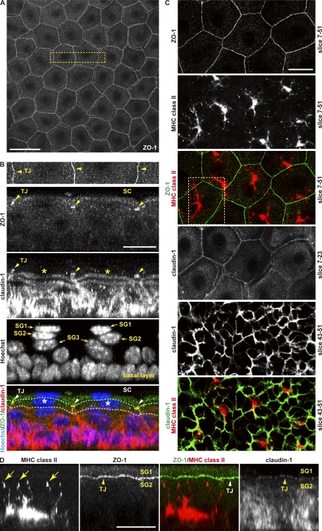
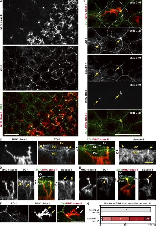
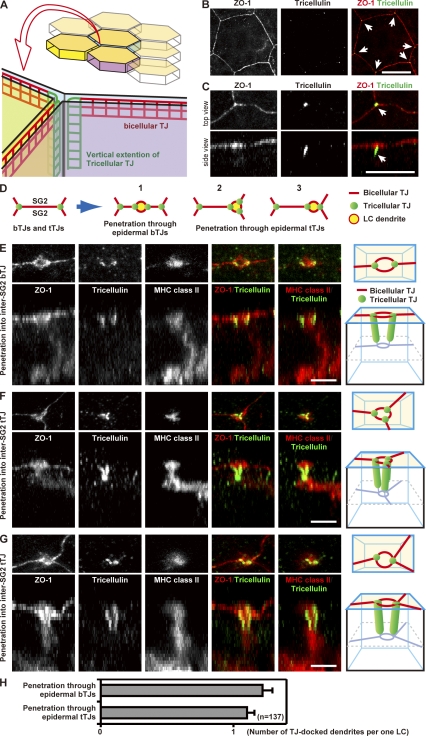
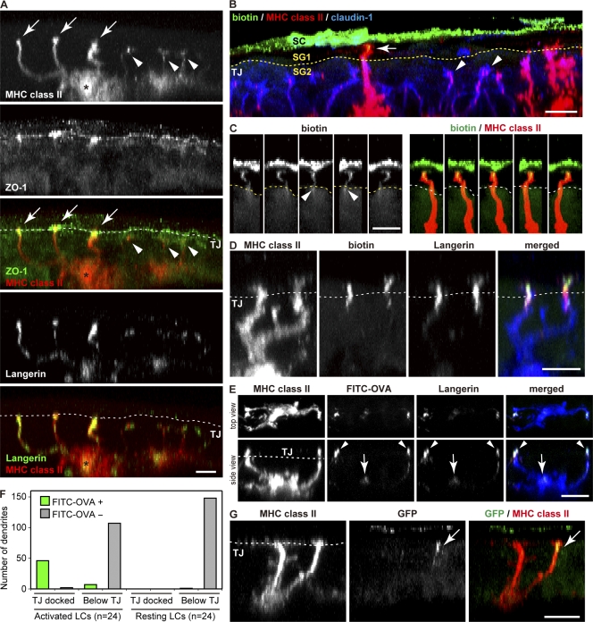
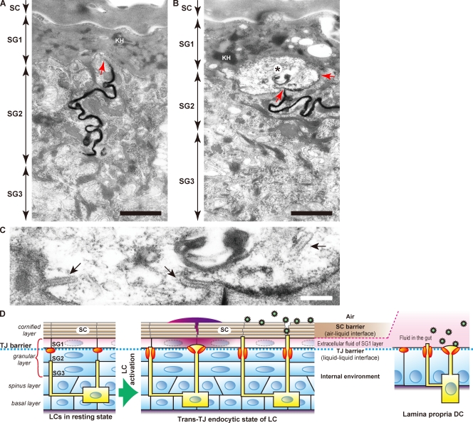
Outermost barriers are critical for terrestrial animals to avoid desiccation and to protect their bodies from foreign insults. Mammalian skin consists of two sets of barriers: stratum corneum (SC) and tight junctions (TJs). How acquisition of external antigens (Ags) by epidermal Langerhans cells (LCs) occur despite these barriers has remained unknown. We show that activation-induced LCs elongate their dendrites to penetrate keratinocyte (KC) TJs and survey the extra-TJ environment located outside of the TJ barrier, just beneath the SC. Penetrated dendrites uptake Ags from the tip where Ags colocalize with langerin/Birbeck granules. TJs at KC-KC contacts allow penetration of LC dendrites by dynamically forming new claudin-dependent bicellular- and tricellulin-dependent tricellular TJs at LC-KC contacts, thereby maintaining TJ integrity during Ag uptake. Thus, covertly under keratinized SC barriers, LCs and KCs demonstrate remarkable cooperation that enables LCs to gain access to external Ags that have violated the SC barrier while concomitantly retaining TJ barriers to protect intra-TJ environment.


Figures

Figure 1.
Figure 1.
3D visualization of epidermal TJs and LCs. (A) TJ network visualized by anti–ZO-1 antibody in epidermal sheet demonstrated en face by confocal microscopy. Bar, 50 µm. (B) A 90° rotation image of the boxed area in A. The top shows an en face image of ZO-1 staining followed by rotation images stained for the indicated TJ molecules. Among the three layers of granular layer, SG1 to SG3, TJs (arrowheads) were found exclusively in the SG2 layer (asterisk and dashed lines), and rotation images display TJs as bright dots with intense ZO-1 and claudin-1 signals (see Video 1). (C) TJs and LCs in unperturbed skin. Z-slice numbers are indicated in each panel. ZO-1 and claudin-1 colocalized at KC–KC TJs, and LCs (MHC II+) coexpressed claudin-1 at high levels. (D) A 90° rotation image of the boxed area in C (see Video 2). Tips of LC dendrites (arrows) localized within TJ barriers (arrowheads). Bars, 20 µm. Data presented is representative of five mice.
Figure 2.
Figure 2.
TJ docking and penetration by activated LCs. (A) Activated LCs exhibit brighter surface MHC II compared with resting LCs. ZO-1high spots identify where LC dendrites dock with TJs. (B) TJ penetration by activated LC dendrites (see Video 2) and accumulation of ZO-1 and claudin-1 at penetration points (arrows). Z-slice numbers are indicated in each panel. (C–F) Various forms of TJ docking and penetration. A 90° rotation of the boxed area in B is shown in C. Dashed lines indicate the SC–SG1 interface. Dendrites penetrated (C, arrows) or docked with (D, arrows) TJs, formed button-like structures (E, arrows), or formed lamellipodia-like protrusions (F, arrows) elongating horizontally between SG1 and SG2 cells after penetrating TJs. Bars: (A and B) 50 µm; (C–F) 10 µm. (G) Numbers of TJ-docked dendrites per cell on resting or activated LC after 12 h of tape stripping. Data presented in AF is representative of five mice, and data in G of three independent experiments.
Figure 3.
Figure 3.
tTJ formation at the TJ penetration point of LC dendrites. (A) Schematic structure of TJs at cell–cell contacts. TJ strands extend vertically below at tricellular contacts to form tTJs (Ikenouchi et al., 2005). (B and C) 3D reconstruction of epidermal sheet stained for ZO-1 and the tTJ protein tricellulin. Tricellulin appears as bright dots (B, arrows) when observed en face, but is found to extend vertically below in 90° rotated images (C, arrows). Bars, 20 µm. (D) Schematic representation of three patterns by which LC dendrites penetrate bTJs or tTJs at the SG2 layer. (E–G) 3D reconstruction images of LC dendrites penetrating TJs as modeled in D (see Video 3). Top rows of each show en face images and bottoms present 90° rotation images accompanied by schematic drawings on the right of each row. Bars, 5 µm. (H) Numbers of bTJ- and tTJ-docked dendrites per LC. The number of dendrites that docked with or penetrated TJs was counted on activated LCs after 12 h of tape stripping. Error bars represent SEM. Data presented in AG is representative of three mice and data in H of three independent experiments.
Figure 4.
Figure 4.
LCs engage in trans-TJ endocytosis via langerin. See Video 4 for more details. (A) Visualization of langerin and MHC II on activated (asterisks) and resting (cell to the right) LCs in relation to TJs as shown by ZO-1 staining. Dashed lines indicate TJs. Langerin accumulated in TJ-docked dendrite tips of activated LCs (arrows). Dendrites of resting LCs (arrowheads) lack langerin accumulation. (B) Biotinylation of LC surface membrane reveals endocytic activity by a TJ-penetrated dendrite (arrow). Dashed lines represent TJs. Dendrites of resting LCs (arrowheads) lack biotin signals. (C) Rotated views of an endocytosing LC dendrite from B. The streak of biotin signal in the dendrite continues within the TJ barrier (arrowheads). (D) Colocalization of biotinylated molecules (green) with langerin (red) in TJ-penetrated dendrites as shown by MHC II (blue) staining. (E) FITC-OVA (green) application demonstrates trans-TJ uptake of Ags by LCs in vivo. FITC-OVA signals colocalized with langerin (red) in TJ-penetrating dendrites (MHC II, blue; arrowheads). Arrows point to accumulation of FITC and langerin signals in perinuclear area of an activated LC. (F) Numbers of FITC-OVA–positive and –negative dendrites. The number of dendrites that docked or did not dock with TJs was counted on resting and activated LCs. (G) GFP-expressing Escherichia coli application demonstrates GFP signal within LC dendrite (arrows). Bars, 10 µm. Data presented in AE and G is representative of three mice and data in F of three independent experiments.
Figure 5.
Figure 5.
Penetrating dendrites and KCs retain TJ barrier function. Electron microscopy images of SC, SG cells, and dendrite tip of an LC. Keratohyalin granules (KH) identify SG1 cells. (A) Lanthanum permeability assay. Intercellular diffusion of lanthanum nitrate (electron-dense streak) is blocked at KC-KC TJs (arrow). (B) TJ-penetrated LC dendrite (asterisk) was found between the SG1 and SG2 cell layers. The junction between the LC dendrite and SG2 cells also inhibited lanthanum diffusion (arrows), suggesting an intact TJ barrier. Bars, 500 nm. (C) Enlarged image of the LC dendrite tip (B, asterisk), in which two of three Birbeck granules (arrows) were observed to generate from the cell membrane. Bar, 100 nm. (D) Schematic model for trans-TJ uptake activity of LCs. Activation of LCs induces elongation of LC dendrites beyond TJ barriers. bTJs and tTJs that are newly formed between LC and SG2 cells preserve the integrity of TJ barriers during this phenomenon. LCs access Ags that have violated the SC barrier in the presence of intact TJ barrier function. Schematics for gut intraepithelial DCs called lamina propria DCs are shown for comparison. Data presented in AC is representative of two mice.

Similar articles

Cited by

References

    1. Aiba S., Katz S.I. 1990. Phenotypic and functional characteristics of in vivo-activated Langerhans cells. J. Immunol. 145:2791–2796 - PubMed
    1. Akhter N., Kobayashi M., Hoshino T. 1993. Avian epidermis contains ATPase- and Ia-positive Langerhans-like cells. Cell Tissue Res. 271:103–106 10.1007/BF00297547 - DOI - PubMed
    1. Brandner J.M., Kief S., Grund C., Rendl M., Houdek P., Kuhn C., Tschachler E., Franke W.W., Moll I. 2002. Organization and formation of the tight junction system in human epidermis and cultured keratinocytes. Eur. J. Cell Biol. 81:253–263 10.1078/0171-9335-00244 - DOI - PubMed
    1. Castell-Rodríguez A.E., Hernández-Peñaloza A., Sampedro-Carrillo E.A., Herrera-Enriquez M.A., Alvarez-Pérez S.J., Rondán-Zarate A. 1999. ATPase and MHC class II molecules co-expression in Rana pipiens dendritic cells. Dev. Comp. Immunol. 23:473–485 10.1016/S0145-305X(99)00031-2 - DOI - PubMed
    1. Enk A.H., Angeloni V.L., Udey M.C., Katz S.I. 1993. An essential role for Langerhans cell-derived IL-1 beta in the initiation of primary immune responses in skin. J. Immunol. 150:3698–3704 - PubMed

Publication types