. 2010 Jan 29;285(5):3126-32.
doi: 10.1074/jbc.M109.055095. Epub 2009 Dec 7.

Proline cis/trans-isomerase Pin1 regulates peroxisome proliferator-activated receptor gamma activity through the direct binding to the activation function-1 domain

Affiliations

Proline cis/trans-isomerase Pin1 regulates peroxisome proliferator-activated receptor gamma activity through the direct binding to the activation function-1 domain

Yoshito Fujimoto et al. J Biol Chem. .

Abstract

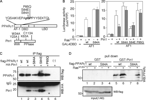
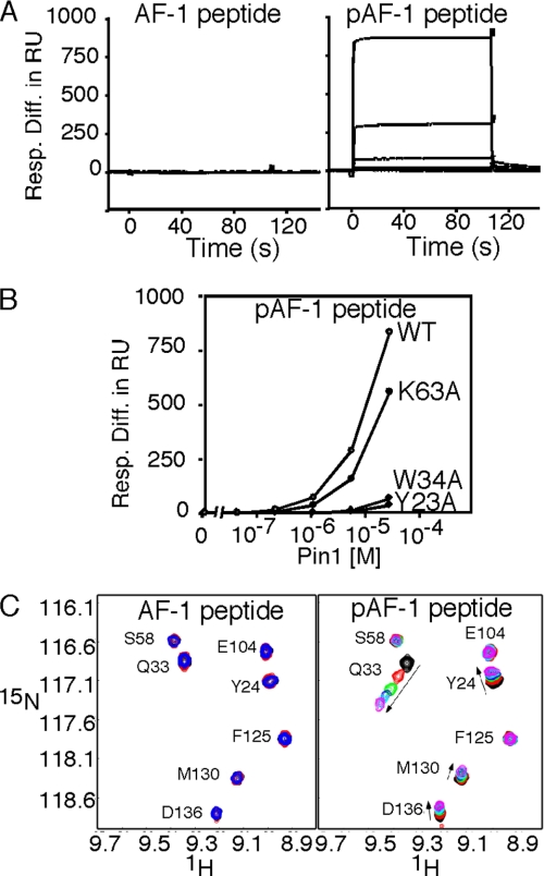
The important roles of a nuclear receptor peroxisome proliferator-activated receptor gamma (PPARgamma) are widely accepted in various biological processes as well as metabolic diseases. Despite the worldwide quest for pharmaceutical manipulation of PPARgamma activity through the ligand-binding domain, very little information about the activation mechanism of the N-terminal activation function-1 (AF-1) domain. Here, we demonstrate the molecular and structural basis of the phosphorylation-dependent regulation of PPARgamma activity by a peptidyl-prolyl isomerase, Pin1. Pin1 interacts with the phosphorylated AF-1 domain, thereby inhibiting the polyubiquitination of PPARgamma. The interaction and inhibition are dependent upon the WW domain of Pin1 but are independent of peptidyl-prolyl cis/trans-isomerase activity. Gene knockdown experiments revealed that Pin1 inhibits the PPARgamma-dependent gene expression in THP-1 macrophage-like cells. Thus, our results suggest that Pin1 regulates macrophage function through the direct binding to the phosphorylated AF-1 domain of PPARgamma.


Figures

FIGURE 1.
FIGURE 1.
Phosphorylation-dependent binding of Pin1 to the AF-1 domain of PPARγ. A, schematic representation of the domain structures of PPARγ and Pin1. The amino acid sequence indicates the AF-1 peptide sequence used for in vitro experiments in this study. The mutated residues in PPARγ are denoted above the sequence. Bold letters indicate the recognition motif of Pin1, in which pS denotes the phosphorylated serine. B, effects of coexpression of RasV12G or Pin1 on the intrinsic activity of the AF-1 domain. GAL4-fused AF-1 domains with the indicated mutations were transfected with or without RasV12G or Pin1 (left and right panels, respectively). Data are presented as the means ± S.D. DBD, DNA-binding domain. C, coimmunoprecipitation of Pin1 with full-length PPARγ in HEK293T cells. Immunoprecipitations (IP) were analyzed by Western blots (WB) probed with either an anti-FLAG or anti-HA antibody (WB:flag and WB:HA, respectively). D, Pin1 interacts with PPARγ in vitro. The cell lysate was incubated with GST or GST-Pin1, and the interaction between Pin1 and PPARγ was detected by Western blotting, using an anti-PPARγ antibody. Cell lysates were also probed with anti-FLAG antibody. LBD, ligand-binding domain.
FIGURE 2.
FIGURE 2.
Phosphorylation-dependent binding of Pin1 to the AF-1 peptide of PPARγ. A, SPR analyses of the interaction between Pin1 and the AF-1 peptide. Traces of the titration of Pin1 on sensor chips immobilized with the non-phosphorylated (AF-1) and phosphorylated AF-1 (pAF-1) peptide (left and right panels, respectively). Concentrations of analyte were 0, 0.04, 0.2, 1, 5, and 25 μm. Data are presented as the response difference in resonance units. B, concentration dependence of the interaction between Pin1 and pAF-1, plotted for each Pin1 protein with the indicated mutation. C, overlay of part of the amide region of the 1H-15N heteronuclear-single-quantum coherence (HSQC) spectra for Pin1 with various amounts of pAF-1 peptide. In the spectra for the pAF-1 titration to Pin1, the overlaid spectra were collected for samples with pAF-1 to Pin1 ratios of 0.0, 0.2, 0.5, 0.7, 1.0, and 1.3. The corresponding spectra for the nonphosphorylated AF-1 with Pin1 and with AF-1 to Pin1 ratios of 0.0, 0.40, and 1.12, are displayed. WT, wild type.
FIGURE 3.
FIGURE 3.
Inhibition of polyubiquitination of PPARγ by Pin1. A, Pin1 inhibits the polyubiquitination of PPARγ in a WW domain-dependent manner. Immunoprecipitations (IP) were analyzed by Western blot (WB) probed with an anti-HA antibody to detect polyubiquitination (bottom panel, WB:HA). Cell lysates were also probed with an anti-FLAG or anti-green fluorescent protein (GFP) antibody (top panel, WB:flag and WB:GFP, respectively). B, Pin1 inhibits the polyubiquitination of PPARγ in a WW domain-dependent manner. Experimental condition was same as A. C, effects of proteasome inhibitor, MG132, on the polyubiquitination of PPARγ. Cells were treated with 10 μm MG132 for 5 h before harvest. Immunoprecipitation condition was the same as in A. wt, wild type; Ub, ubiquitin.
FIGURE 4.
FIGURE 4.
Inhibitory effect of Pin1 on PPARγ-dependent transcription. A, schematic representation of the human FABP4 5′ upstream region and a series of DNA constructs used for the reporter assay. PPRE was found at −5216 bp (arrowhead) within the mammalian conserved region. The figure was drawn using the ENCODE web server. B, PPARγ-dependent activation of the human FABP4 enhancer. The indicated reporter plasmids, PPARγ1 and RXRα genes were cotransfected into HEK293T cells, and the transcriptional activities were determined by the luciferase activity in the presence or absence of 0.5 μm BRL49653. C, effect of Pin1 on the PPARγ-dependent activation of the FABP4 promoter. The indicated reporter plasmids, PPARγ1 and RXRα genes were cotransfected into HEK293T cells with or without Pin1. D, effects of Pin1 mutations on the Pin1-mediated inhibition of the PPARγ-dependent transcription. The indicated reporter plasmids, PPARγ1 and RXRα genes were cotransfected into HEK293T cells with or without Pin1 carrying indicated mutations. E, effects of MG132 on the PPARγ-dependent transcription. The indicated reporter plasmids and PPARγ1 and RXRα genes were co-transfected into HEK293T cells. After 12 h, cells were treated with or without 10 μm MG132 for another 6 h. Luciferase activity was not normalized by any internal control because MG132 might change the stability of the control protein. F, effect of the Pin1 knockdown on the PPARγ-dependent up-regulation of endogenous proteins in THP-1 cells. Cells were differentiated into macrophage-like cells by 5 nm phorbol 12-myristate 13-acetate. After 24 h, cells were treated with 0.5 μm BRL49653 for additional 24 h. Cellular lysates including 50 μg proteins were separated by SDS-PAGE, and protein expression was analyzed by Western blots probed with either an anti-FABP4, anti-HO-1 or anti-β-Tubulin antibody. G, fluorescence-activated cell sorter analysis of the effect of the Pin1 knockdown on the PPARγ-dependent CD36 expression in THP-1 cells. Cells were differentiated into macrophage-like cells by 5 nm phorbol 12-myristate 13-acetate. After 24 h, cells were treated with 0.5 μm BRL49653 for additional 24 h. 0.5 × 10⁶ cells were incubated with fluorescein isothiocyanate (FITC)-labeled anti-CD36 antibody, and the fluorescent intensity was analyzed by fluorescence-activated cell sorter. wt, wild type; DMSO, dimethyl sulfoxide.
FIGURE 5.
FIGURE 5.
Model of the Pin1-mediated regulation of PPARγ activity. Binary switch model of the Pin1-mediated regulation of PPARγ. In the absence of phosphorylation signals, PPARγ is polyubiquitinated. Ras-mediated kinase signals induce the phosphorylation of Ser84. Binding of Pin1 to the phosphorylated AF-1 prevents the polyubiquitination of PPARγ, resulting in slow turnover of the PPARγ protein. Because the proteasome inhibitor, MG132, reduces PPARγ activity, the efficient transcription seems to require continuous turnover of PPARγ through the ubiquitin-proteasome pathway. Then, Pin1-mediated inhibition of polyubiquitination results in the reduction of the PPARγ activity. In this regulation, binding through the WW domain is sufficient for the inhibition of polyubiquitination, and the PPIase activity of Pin1 is dispensable. LBD, ligand-binding domain; DBD, DNA-binding domain; (Ub)n, polyubiquitination.

References

    1. Lee C. H., Olson P., Evans R. M. (2003) Endocrinology 144, 2201–2207 - PubMed
    1. Rosen E. D., Spiegelman B. M. (2001) J. Biol. Chem. 276, 37731–37734 - PubMed
    1. Mukherjee R., Jow L., Croston G. E., Paterniti J. R., Jr. (1997) J. Biol. Chem. 272, 8071–8076 - PubMed
    1. Waku T, Shiraki T, Oyama T, Fujimoto Y, Maebara K, Kamiya N, Jingami H, Morikawa K. (2009) J. Mol. Biol. 385, 188–199 - PubMed
    1. Glass C. K., Rosenfeld M. G. (2000) Genes Dev. 14, 121–141 - PubMed

Publication types

LinkOut - more resources