

An alternative form of replication protein a expressed in normal human tissues supports DNA repair
- PMID: 19996105
- PMCID: PMC2836084
- DOI: 10.1074/jbc.M109.079418
An alternative form of replication protein a expressed in normal human tissues supports DNA repair
Abstract
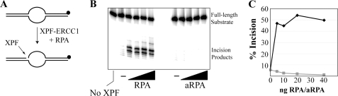
Replication protein A (RPA) is a heterotrimeric protein complex required for a large number of DNA metabolic processes, including DNA replication and repair. An alternative form of RPA (aRPA) has been described in which the RPA2 subunit (the 32-kDa subunit of RPA and product of the RPA2 gene) of canonical RPA is replaced by a homologous subunit, RPA4. The normal function of aRPA is not known; however, previous studies have shown that it does not support DNA replication in vitro or S-phase progression in vivo. In this work, we show that the RPA4 gene is expressed in normal human tissues and that its expression is decreased in cancerous tissues. To determine whether aRPA plays a role in cellular physiology, we investigated its role in DNA repair. aRPA interacted with both Rad52 and Rad51 and stimulated Rad51 strand exchange. We also showed that, by using a reconstituted reaction, aRPA can support the dual incision/excision reaction of nucleotide excision repair. aRPA is less efficient in nucleotide excision repair than canonical RPA, showing reduced interactions with the repair factor XPA and no stimulation of XPF-ERCC1 endonuclease activity. In contrast, aRPA exhibits higher affinity for damaged DNA than canonical RPA, which may explain its ability to substitute for RPA in the excision step of nucleotide excision repair. Our findings provide the first direct evidence for the function of aRPA in human DNA metabolism and support a model for aRPA functioning in chromosome maintenance functions in nonproliferating cells.
Figures


Similar articles
-
Functions of alternative replication protein A in initiation and elongation.Biochemistry. 2010 Jul 20;49(28):5919-28. doi: 10.1021/bi100380n. Biochemistry. 2010. PMID: 20545304 Free PMC article.
-
Rad52-mediated DNA annealing after Rad51-mediated DNA strand exchange promotes second ssDNA capture.EMBO J. 2006 Nov 29;25(23):5539-48. doi: 10.1038/sj.emboj.7601412. Epub 2006 Nov 9. EMBO J. 2006. PMID: 17093500 Free PMC article.
-
Dynamic regulatory interactions of rad51, rad52, and replication protein-a in recombination intermediates.J Mol Biol. 2009 Jul 3;390(1):45-55. doi: 10.1016/j.jmb.2009.05.009. Epub 2009 May 13. J Mol Biol. 2009. PMID: 19445949
-
Replication protein A, the laxative that keeps DNA regular: The importance of RPA phosphorylation in maintaining genome stability.Semin Cell Dev Biol. 2019 Feb;86:112-120. doi: 10.1016/j.semcdb.2018.04.005. Epub 2018 Nov 13. Semin Cell Dev Biol. 2019. PMID: 29665433 Review.
-
[DNA homologous recombination repair in mammalian cells].Postepy Biochem. 2006;52(2):180-93. Postepy Biochem. 2006. PMID: 17078508 Review. Polish.
Cited by
-
Genome Stability Maintenance in Naked Mole-Rat.Acta Naturae. 2017 Oct-Dec;9(4):31-41. Acta Naturae. 2017. PMID: 29340215 Free PMC article.
-
The DNA damage response kinases DNA-dependent protein kinase (DNA-PK) and ataxia telangiectasia mutated (ATM) Are stimulated by bulky adduct-containing DNA.J Biol Chem. 2011 Jun 3;286(22):19237-46. doi: 10.1074/jbc.M111.235036. Epub 2011 Apr 12. J Biol Chem. 2011. PMID: 21487018 Free PMC article.
-
ATRIP from TopBP1 to ATR--in vitro activation of a DNA damage checkpoint.Proc Natl Acad Sci U S A. 2010 Aug 3;107(31):13561-2. doi: 10.1073/pnas.1008909107. Epub 2010 Jul 26. Proc Natl Acad Sci U S A. 2010. PMID: 20660767 Free PMC article. No abstract available.
-
Dynamic elements of replication protein A at the crossroads of DNA replication, recombination, and repair.Crit Rev Biochem Mol Biol. 2020 Oct;55(5):482-507. doi: 10.1080/10409238.2020.1813070. Epub 2020 Aug 28. Crit Rev Biochem Mol Biol. 2020. PMID: 32856505 Free PMC article. Review.
-
Tipin-replication protein A interaction mediates Chk1 phosphorylation by ATR in response to genotoxic stress.J Biol Chem. 2010 May 28;285(22):16562-71. doi: 10.1074/jbc.M110.110304. Epub 2010 Mar 15. J Biol Chem. 2010. PMID: 20233725 Free PMC article.
References
Publication types
MeSH terms
Substances
Grants and funding
LinkOut - more resources
Full Text Sources
Other Literature Sources
Molecular Biology Databases
Research Materials
