. 2010 Feb 19;285(8):5614-23.
doi: 10.1074/jbc.M109.073197. Epub 2009 Dec 7.

Huntingtin-associated protein-1 interacts with pro-brain-derived neurotrophic factor and mediates its transport and release

Affiliations

Huntingtin-associated protein-1 interacts with pro-brain-derived neurotrophic factor and mediates its transport and release

Linda Lin-yan Wu et al. J Biol Chem. .

Abstract

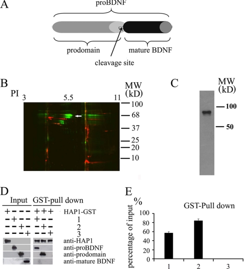
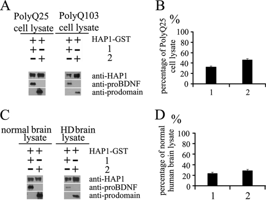
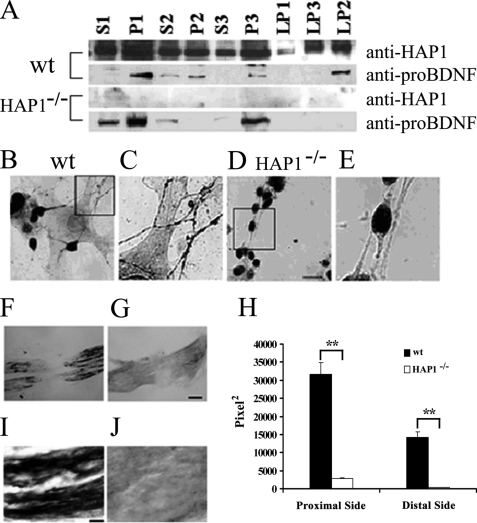
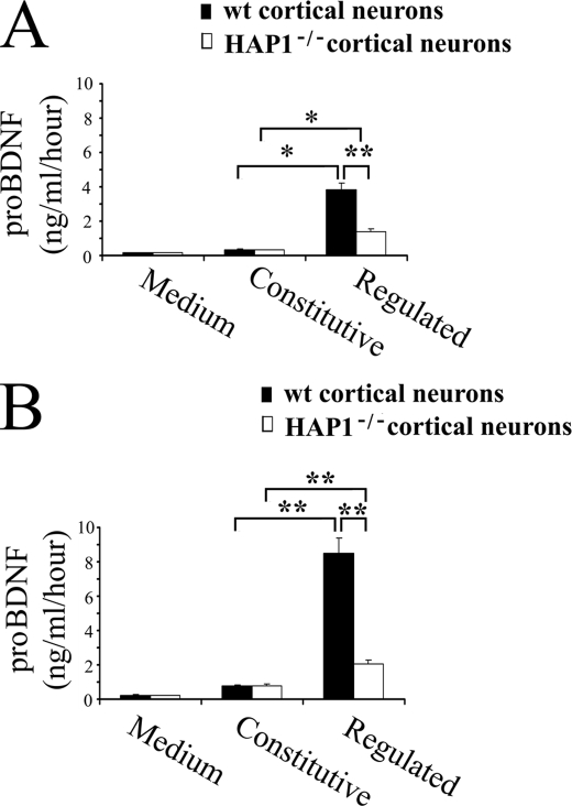
Brain-derived neurotrophic factor (BDNF) plays a pivotal role in brain development and synaptic plasticity. It is synthesized as a precursor (pro-BDNF), sorted into the secretory pathway, transported along dendrites and axons, and released in an activity-dependent manner. Mutant Huntingtin with expanded polyglutamine (polyQ) and the V66M polymorphism of BDNF reduce the dendritic distribution and axonal transport of BDNF. However, the mechanism underlying this defective transport remains unclear. Here, we report that Huntingtin-associated protein-1 (HAP1) interacts with the prodomain of BDNF and that the interaction was reduced in the presence of polyQ-expanded Huntingtin and BDNF V66M. Consistently, there was reduced coimmunoprecipitation of pro-BDNF with HAP1 in the brain homogenate of Huntington disease. Pro-BDNF distribution in the neuronal processes and its accumulation in the proximal and distal segments of crushed sciatic nerve and the activity-dependent release of pro-BDNF were abolished in HAP1(-/-) mice. These results suggest that HAP1 may participate in axonal transport and activity-dependent release of pro-BDNF by interacting with the BDNF prodomain. Accordingly, the decreased interaction between HAP1 and pro-BDNF in Huntington disease may reduce the release and transport of BDNF.


Figures

FIGURE 1.
FIGURE 1.
Association of HAP1 with pro-BDNF and the prodomain in vitro. A, domain structure of pro-BDNF, prodomain, and mature BDNF. Pro-BDNF is the unprocessed full-length form after synthesis and is cleaved to produce mature BDNF. The prodomain is the fragment of 1–130 amino acids of pro-BDNF. B, binding proteins were collected by passing rat brain lysate through affinity columns containing the recombinant prodomain and mature BDNF. DIGE two-dimensional gel was used to reveal all proteins bound to the recombinant prodomain (green) and mature BDNF (red) affinity columns. When sequenced by MALDI-TOF, one of these proteins was shown to be HAP1 (white arrow). PI, isoelectric point. C, affinity-purified prodomain-binding proteins was probed with rabbit anti-HAP1 antibody. The antibody recognized a band (around 75 kDa) in the prodomain-binding proteins. D, HAP1 GST fusion proteins were immobilized on glutathione-agarose beads and incubated with pro-BDNF (1), prodomain (2), and mature BDNF (3). Bound proteins were detected by Western blotting with the indicated antibodies. E, analysis of percentage of pulldown proteins. The staining intensity of each band on the blots in D was quantified by the ImageJ program (National Institutes of Health). The percentage of input was calculated using input proteins as 100%. The data are presented as mean ± S.E. (n = 3).
FIGURE 2.
FIGURE 2.
Mutant htt in PC12 cell lysate and HD brain lysate decrease the efficiency of the interaction of HAP1 with pro-BDNF. HAP1 GST fusion protein was immobilized on glutathione-agarose beads and incubated with pro-BDNF (1) and the prodomain (2) in the presence of polyQ25- or polyQ103-containing Htt-transfected PC12 cell lysate or in the presence of normal or HD human brain lysate. Binding proteins were detected by immunoblotting. A, quantity of proteins pulled down in the presence of polyQ25-htt PC12 cell lysate or polyQ103-htt PC12 cell lysate is shown in the blot. B, percentages of proteins in the presence of polyQ103-htt PC12 cell lysate were calculated using the polyQ25-htt PC12 cell lysate as 100% references. C, quantity of proteins pulled down in the presence of normal brain lysate or HD brain lysate is shown in the blot. D, percentages of proteins in the presence of HD brain lysate were calculated using normal brain lysate as 100%. The blot intensity of each band was quantified by the NIH ImageJ program. The data are presented as mean ± S.E., from three separate experiments.
FIGURE 3.
FIGURE 3.
HAP1-pro-BDNF complex is altered in HD. A, rabbit anti-HAP1 antibody was immobilized on protein A beads and incubated with normal human brain or HD brain lysates. Blots were detected with anti-pro-BDNF, anti-mature BDNF, anti-sortilin, or anti-pro-BDNF plus the immunizing peptide, or anti-HAP antibodies. Lane 1, prodomain standard; lane 2, mature BDNF standard; lane 3, HAP1 immunoprecipitation from HD human brain; lane 4, HAP1 immunoprecipitation from normal human brain. B, percentages of relative protein staining intensity of HD human brain lysate compared with normal human brain lysate (the area of particles that stands for the relative staining intensity was quantified by the ImageJ program). The data are presented means ± S.E. of three separate experiments.
FIGURE 4.
FIGURE 4.
Effect of the V66M mutation on the interaction between HAP1-A and BDNF prodomain. A, input of the WT prodomain and V66M prodomain is shown in panel a. HAP1 GST fusion proteins were immobilized on glutathione-agarose beads and incubated with the WT prodomain or the V66M prodomain (panel b). In separate reaction vials, the reactions as described in panel b were carried in the presence of polyQ25- (panel c) or polyQ103 Htt (panel d)-transfected PC12 cell lysate or in the presence of normal (panel e) or Huntington disease (panel f) human brain lysate. The quantities of binding proteins were detected by immunoblotting as indicated. B, quantification of the percentage of proteins pulled down at different conditions. Bar 1(panels b/a), comparison of the percentages of the WT and V66M prodomains; bar 2 (panels d/c), the comparison of the percentages of WT and V66M prodomains pulled down in the presence of polyQ103 PC12 cell lysate; bar 3 (panels f/e), comparison of the percentages of WT and V66M prodomains pulled down in the presence of HD brain lysate. The staining intensity of each band was quantified by the ImageJ program, and data are presented as means ± S.E. of three separate experiments.
FIGURE 5.
FIGURE 5.
Colocalization of HAP1-A with BDNF subdomains in PC12 cells. A, PC12 cells were transfected with plasmids HAP1-A/EGFP (green) in combination with either full-length pro-BDNF-Ds-Red (row 1) or prodomain-Ds-Red (2) (row 2) (red), respectively. Confocal images were taken and merged (yellow). Yellow dots indicate vesicles with both HAP1-A and BDNF subdomains. The snapshot represents the rate of colocalization quantitatively analyzed by the quantitative analysis software on a Leica SP5 confocal microscope. B, quantitative data showed pro-BDNF and prodomain highly colocalized with HAP1-A. The data are presented as mean ± S.E. (n = 20). C, double labeling of pro-BDNF and HAP1 in cultured cortical neurons from WT and HAP1−/− mice. Pro-BDNF is stained in red and HAP1 in green. No HAP1 was stained in HAP1−/− neurons. Pro-BDNF and HAP1 are highly colocalized in the cytoplasm and neuritis in WT neurons. In HAP1−/− neurons, pro-BDNF is only present in soma. Phase contrast images showed cell bodies and neurites of WT and HAP1−/− neurons.
FIGURE 6.
FIGURE 6.
HAP1 is required for the transport of pro-BDNF. A, subcellular fractions of sucrose gradient from P1 neonatal wild type (wt) and HAP1−/− mouse cortex were detected by Western blot using sheep anti-pro-BDNF and rabbit anti-HAP1. Equal amounts of protein (65 μg) were loaded in each lane. B, pro-BDNF immunohistochemistry of primary cultured cortical neurons from P1 neonatal WT mouse. C, higher magnification view of the field highlighted in B. D, pro-BDNF immunohistochemistry of primary cultured cortical neurons from P1 neonatal HAP1−/− mouse (scale bar, 15 μm). E, higher magnification view of the field highlighted in D. F, photomicrograph of a crushed sciatic nerve section stained for pro-BDNF from a P1 neonatal WT mouse. G, photomicrograph of a crushed sciatic nerve section stained for pro-BDNF from a P1 neonatal HAP1−/− mouse. H, quantitative data on the relative staining of pro-BDNF in the sciatic nerve. y axis stands for the relative amount quantified by the ImageJ program. The data are presented as mean ± S.E., n = 3. **, p < 0.01, Student's t test, scale bar, 15 μm. I, high magnification of micrograph of the proximal segment of crushed nerve stained for pro-BDNF from a P1 neonatal WT mouse; scale bar, 60 μm. J, high magnification of micrograph of the proximal segment of crushed nerve stained for pro-BDNF from a P1 neonatal HAP1−/− mouse.
FIGURE 7.
FIGURE 7.
Role of HAP1 in the constitutive and activity-dependent secretion of pro-BDNF from culture cortical neurons. A, experiment was performed on naive cortical neurons cultured for 48 h. Neurons were conducted under constitutive and depolarization secretion conditions. All collected media were subjected for quantification of pro-BDNF with an ELISA developed in our laboratory. B, experiment was performed in the same conditions as above except that neurons were transfected with the pro-BDNF plasmid. All results are presented as a mean ± S.E. determined from analysis of six independent experiments (*, p < 0.05; **, p < 0.01, n = 6, Student's t test).

Similar articles

Cited by

References

    1. Chao M. V. (1992) Neuron 9, 583–593 - PubMed
    1. Goodman L. J., Valverde J., Lim F., Geschwind M. D., Federoff H. J., Geller A. I., Hefti F. (1996) Mol. Cell. Neurosci. 7, 222–238 - PubMed
    1. Huang E. J., Reichardt L. F. (2001) Annu. Rev. Neurosci. 24, 677–736 - PMC - PubMed
    1. Lu B. (2003) Neuron 39, 735–738 - PubMed
    1. Poo M. M. (2001) Nat. Rev. Neurosci. 2, 24–32 - PubMed

Publication types

MeSH terms