. 2009 Dec 22;106(51):21666-71.
doi: 10.1073/pnas.0912180106. Epub 2009 Dec 8.

Gli2 trafficking links Hedgehog-dependent activation of Smoothened in the primary cilium to transcriptional activation in the nucleus

Affiliations

Gli2 trafficking links Hedgehog-dependent activation of Smoothened in the primary cilium to transcriptional activation in the nucleus

Jynho Kim et al. Proc Natl Acad Sci U S A. .

Abstract

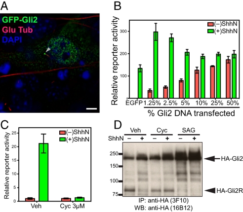
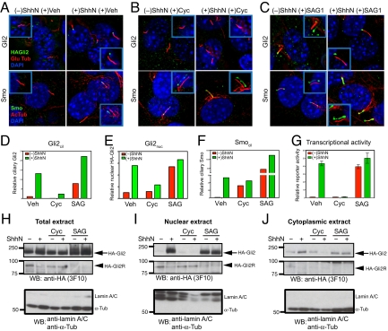
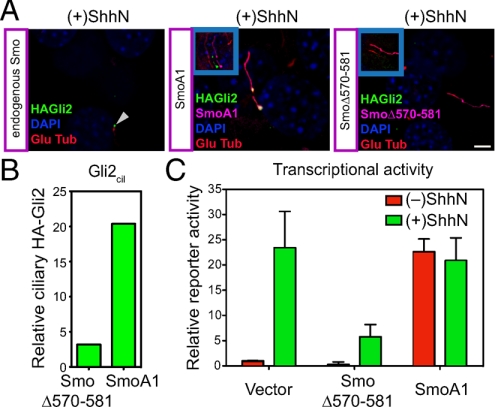
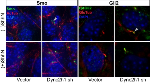
Stimulation by the extracellular Hedgehog (Hh) protein signal has been shown to alter ciliary localization of the mammalian Hh receptor components Smoothened (Smo) and Patched (Ptc), and mutations that disrupt the structure and function of the cilium also disrupt Hh-induced changes in gene expression. But how ciliary events affect gene expression in the nucleus is not known, and to address this question we have characterized the cellular trafficking of Gli2, the principal mediator of Hh-dependent transcriptional activation. From a combination of pharmacological and genetic manipulations we find in resting cells that both Gli2 and Smo appear to shuttle in and out of the cilium, with Gli2 but not Smo requiring intact cytoplasmic microtubules for ciliary entry and both requiring the ciliary retrograde motor, cytoplasmic dynein 2, for ciliary exit. We also find that changes in ciliary and nuclear trafficking of Gli2 are triggered by the Hh-dependent accumulation of activated Smo in the cilium, resulting in a shift from primarily cytoplasmic localization to accumulation at the distal tip of the cilium and within the nucleus. Gli2 thus functions as a dynamic monitor of Smo activity in the cilium and thereby links Hh pathway activation in the cilium to transcriptional activation in the nucleus.


Conflict of interest statement

The authors declare no conflict of interest.

Figures

Fig. 1.
Fig. 1.
Physiological regulation of exogenously introduced Gli2. (A) NIH 3T3 cells were transiently transfected for expression of GFP-Gli2 and stained 48 h later. Overexpressed Gli2 is localized in the cytoplasm, at one end of the primary cilium (visualized with anti-detyrosinated tubulin), and in the nucleus (visualized with DAPI). (Scale bar, 5 μm.) (B) NIH 3T3 cells transfected for expression of Gli2 show that high levels produce unregulated reporter expression irrespective of ShhN stimulation, whereas lower levels can contribute to increased reporter expression with regulation by ShhN. Gli-luciferase reporter and control SV40-Renilla luciferase were cotransfected with the indicated proportion of Gli2 DNA (% of total DNA, wt/wt). Following transfection, cells were grown to confluency, incubated with Shhn, and assayed for reporter activity. Error bars indicate SD. (C) A Hh-responsive NIH 3T3/HA-Gli2 clone. An NIH 3T3 cell clone with a stably integrated construct for expression of HA-Gli2 was established and chosen for its low level of HA-Gli2 expression and assayed with Gli-luciferase reporter for response to ShhN stimulation. This clone displays Hh-responsive, cyclopamine-suppressible pathway activity. Error bars indicate SD. (D) 3T3/HA-Gli2 cells were incubated in the absence or presence of ShhN with vehicle (Veh), cyclopamine (Cyc), or SAG1, and then lysed and analyzed by IP-Western with anti-HA antibodies. Markers of molecular mass are indicated on the Left, and migration of full-length Gli2 (HA-Gli2, arrow) and its repressor form (HA-Gli2R, arrowhead) on the Right.
Fig. 2.
Fig. 2.
Gli2 accumulate at the primary cilium and the nucleus upon Hh stimulation, and pharmacological manipulation of Smo activity in cilia affects ciliary and nuclear accumulation and transcriptional activity of Gli2. (A–F) NIH 3T3/HA-Gli2 cells were incubated with or without ShhN in the presence of vehicle control (Veh), cyclopamine (3 μM) or SAG1 (400 nM) for 24 h. Cells were then stained to visualize Gli2, Smo, the primary cilium, and the nucleus as indicated. The Insets show shifted overlays. (A) ShhN-induced accumulation of Gli2 and Smo at the primary cilium. NIH 3T3/HA-Gli2 cells were grown to confluency and incubated in the presence or absence of ShhN; cells were stained to visualize HA-Gli2, the primary cilium (Glu tubulin), and the nucleus (DAPI) in Upper panels, or Smo, the primary cilium (acetylated tubulin), and the nucleus (DAPI) in Lower panels. (B) Cyclopamine induces Smo accumulation in the primary cilium but inhibits that of Gli2. (C) Smo agonist SAG1 induces accumulation of Gli2 and Smo in the cilium. (D–F) The amount of Gli2 in cilia (Gli2cil) and Gli2 in nuclei (Gli2nuc), Smo in cilia (Smocil) were quantified from complete Z-series of immunofluorescence images. (G) Gli-luciferase reporter activity was assayed in parallel. Error bars indicate SD. (H–J) Total extract, nuclear and cytoplasmic fractions were prepared from cells grown in parallel with A–F and analyzed by immunoblotting with anti-HA antibody. The Upper and Middle panels show full-length HA-Gli2 (HA-Gli2, arrow) and its repressor form (HA-Gli2R, arrowhead), respectively. Note the reciprocal accumulation of HA-Gli2 and HA-Gli2R in the absence or presence of ShhN in the nuclear extract. In the Lower panel, immunoblotting of the same extracts with a mixture of antibodies specific to lamin A/C and α-tubulin shows that only the nuclear extracts contain lamin A/C, and only the cytoplasmic extracts contain α-tubulin. Note that the total extract shows a strong α-tubulin band and also a faint lamin A/C band.
Fig. 3.
Fig. 3.
Mutational manipulation of Smo activity in cilia affects ciliary accumulation and transcriptional activity of Gli2. (A) NIH 3T3/HA-Gli2 cells were transfected with constructs for expression of mutant activated Smo (SmoA1) or a mutant dominant-negative Smo (SmoΔ570–581) (see Fig. S4). Cells were incubated with ShhN and stained for Smo and Gli2. As an internal control, the Left panel shows a cilium (arrowhead) from an untransfected cell (containing only endogenous Smo) in a SmoΔ570–581 transfection experiment. The Insets show shifted overlays. (B) The levels of ciliary Gli2 (Gli2cil) in complete Z-series of immunofluorescence images for cells transfected either with dominant-negative Smo (SmoΔ570–581) or activated Smo (SmoA1) in the presence of ShhN are shown. (C) Gli-luciferase reporter activity was assayed in parallel. Error bars indicate SD.
Fig. 4.
Fig. 4.
Gli2 shuttles in and out of cilia. NIH 3T3/HA-Gli2 cells infected with viruses from the LMP shRNA control vector or the same vector carrying cytoplasmic dynein 2 heavy chain (Dync2h1) shRNA were selected for puromycin resistance. Cells grown to confluency were incubated in the presence or absence of ShhN and stained for Smo or Gli2 as indicated. For greater clarity, Smo staining (green) in the Left four panels is shown as a shifted overlay with acetylated tubulin (red). Note the accumulation of both Smo and Gli2 in the primary cilium (white arrowheads) of cells infected with Dync2h1 shRNA viruses, even in the absence of ShhN, showing that impairment of the retrograde motor cytoplasmic dynein 2 causes ciliary accumulation of Smo and Gli2 in the primary cilium.
Fig. 5.
Fig. 5.
Microtubules are required for Gli2 but not Smo entry into cilia. (A) The microtubule inhibitor vinblastine (VNB) blocks ShhN-induced Gli2 accumulation in the primary cilium, but does not inhibit ciliary Smo accumulation. Cells incubated in the absence or presence of ShhN, or with 100 nM of VNB in the presence of ShhN for 6 h were stained for HA-Gli2 (Left panels) and Smo (Right panels). The Insets show shifted overlays. (Scale bar, 5 μm.) (B) The levels of Gli2 in cilia (Gli2cil) quantified from complete Z-series of immunofluorescence images are shown for cells incubated for 6 h with ShhN and increasing concentrations of VNB from 12.5 nM to 200 nM. (C) The levels of Smo in cilia (Smocil) in cells treated with ShhN and vinblastine were similarly determined. (D) Gli-luciferase reporter activity after 6 h of ShhN treatment in the presence of known pathway inhibitors (Cyc 3 μM, SANT-1 100 nM), VNB (100 nM), and an agonist SAG1 (400 nM). Note that 6 h of ShhN stimulation produces reduced but nevertheless measurable reporter activity, which is completely inhibited by VNB treatment at 100 nM. Error bars indicate SD.

Similar articles

Cited by

References

    1. Chiang C, et al. Cyclopia and defective axial patterning in mice lacking Sonic hedgehog gene function. Nature. 1996;383:407–413. - PubMed
    1. Nusslein-Volhard C, Wieschaus E. Mutations affecting segment number and polarity in Drosophila. Nature. 1980;287:795–801. - PubMed
    1. Jessell TM. Neuronal specification in the spinal cord: Inductive signals and transcriptional codes. Nat Rev Genet. 2000;1:20–29. - PubMed
    1. Ingham PW, McMahon AP. Hedgehog signaling in animal development: Paradigms and principles. Genes Dev. 2001;15:3059–3087. - PubMed
    1. Zhao C, et al. Hedgehog signalling is essential for maintenance of cancer stem cells in myeloid leukaemia. Nature. 2009;458:776–779. - PMC - PubMed

Publication types

LinkOut - more resources