. 2009 Dec 15;15(24):7593-7601.
doi: 10.1158/1078-0432.CCR-09-2317.

Inhibition of Functional Hyaluronan-CD44 Interactions in CD133-positive Primary Human Ovarian Carcinoma Cells by Small Hyaluronan Oligosaccharides

Affiliations

Inhibition of Functional Hyaluronan-CD44 Interactions in CD133-positive Primary Human Ovarian Carcinoma Cells by Small Hyaluronan Oligosaccharides

Mark G Slomiany et al. Clin Cancer Res. .

Abstract

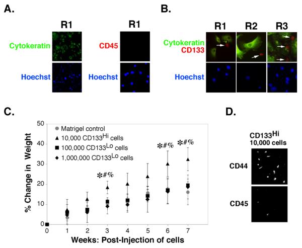
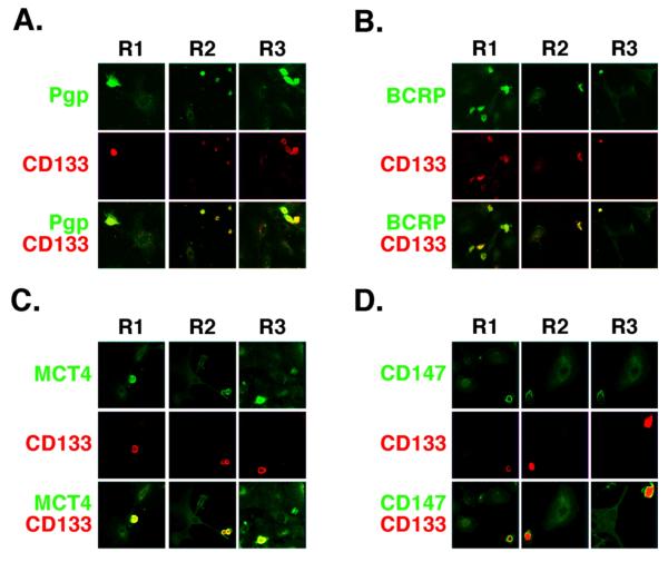
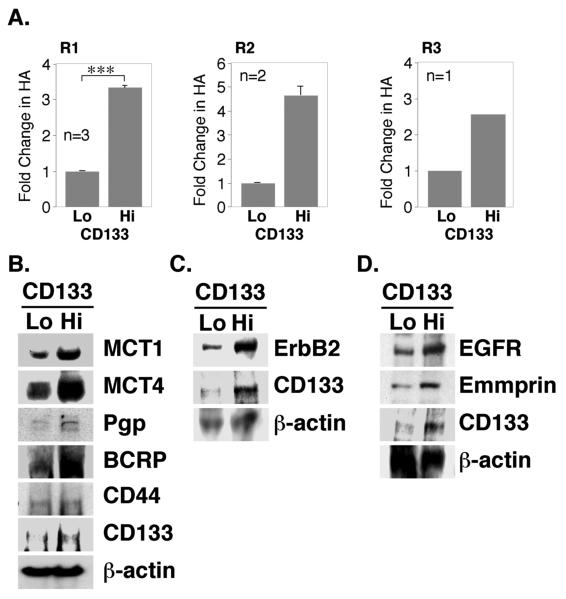
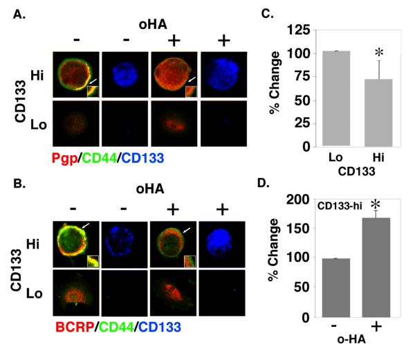
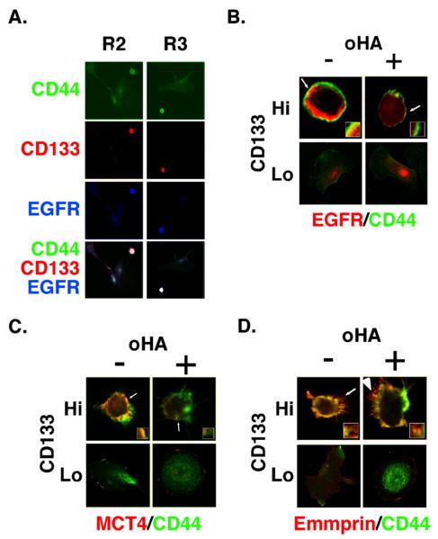
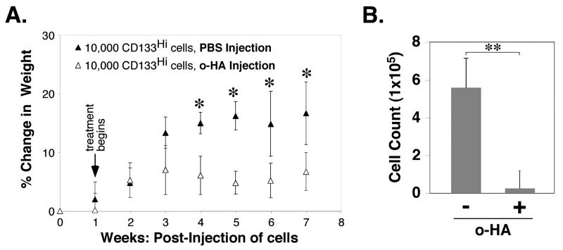
PURPOSE: CD44 is one of the most common markers used for identification of highly tumorigenic subpopulations of human carcinoma cells, but little is known about the function of CD44 or its major ligand, hyaluronan, in these cells. The purpose of this study was to investigate the involvement of hyaluronan and its interaction with CD44 in the properties of a tumorigenic subpopulation of primary ovarian carcinoma cells. EXPERIMENTAL DESIGN: A tumorigenic subpopulation was identified in ascites fluids from ovarian carcinoma patients by expression of high CD133 levels. Treatment with small hyaluronan oligosaccharides, which dissociate constitutive hyaluronan polymer-CD44 interactions, was used to test the importance of hyaluronan-CD44 interaction in assembly of multidrug and monocarboxylate transporters and receptor tyrosine kinases in the plasma membrane of cells with high CD133 levels, and in the tumorigenic capacity of the CD133-high subpopulation. RESULTS: Although total CD44 levels were similar in cells with high or low CD133 expression, CD44 was present in close association with transporters, receptor tyrosine kinases, and emmprin (CD147) in the plasma membrane of cells with high CD133 levels. Treatment with small hyaluronan oligosaccharides reduced association of the transporters and receptor tyrosine kinases with CD44 in the plasma membrane, diminished drug transporter activity, and inhibited i.p. tumorigenesis in these cells. CONCLUSIONS: We conclude that hyaluronan-CD44 interaction plays an important role in the properties of highly tumorigenic cells by stabilizing oncogenic complexes in their plasma membrane, and that treatment with hyaluronan-CD44 antagonists provides a logical therapeutic approach for abrogating the properties of these cells. (Clin Cancer Res 2009;15(24):7593-601).


Figures

FIGURE 1
FIGURE 1. Identification of a CD133-hi tumorigenic sub-population of carcinoma cells in patient ascites
A,B. Unsorted ovarian carcinoma cells from patients R1, R2 or R3 were plated on chamber slides for 24h and processed for immuno-staining as indicated. Cells were visualized by epifluorescence. Arrows in B indicate CD133-hi cells. C, D. Carcinoma cells from patient R1, magnetically sorted for CD133, were injected into the peritoneum of SCID mice as indicated. C. Tumor growth after injection of 104 CD133-hi cells in Matrigel was compared to tumor growth after injection of 105 or 106 CD133-lo cells in Matrigel (or Matrigel alone, as control), as assessed by percent change in weight due to ascites accumulation [(weight at week n/weight at day 0)-1]*100. Significant differences were found between CD133-hi @ 104 cells and: CD133-lo @ 105 cells (*p<.05), CD133-lo @ 106 cells (#p<.05), or Matrigel control (%p<.05), as determined by one-way ANOVA with Bonferroni comparison of significant differences between points. D. After 7 weeks, ascites was drawn from the peritoneum of animals receiving CD133-hi cells and cells plated for 24h before staining for CD44 and CD45. Ascites was absent from animals that received CD133-lo cells or Matrigel alone and significant numbers of cells could not be retrieved from these animals.
FIGURE 2
FIGURE 2. Co-distribution of signaling and transporter proteins in CD133-hi primary ovarian carcinoma cells
A-D. Low power confocal images of unsorted ovarian carcinoma cells from patients R1, R2 and R3 that were plated on chamber slides for 24h and stained for CD133 (red) and either Pgp (A), BCRP (B), MCT4 (C), or emmprin/CD147 (D) (green). Most cells lacked significant staining for CD133 (CD133-lo cells); these cells showed relatively low levels of Pgp, BCRP, MCT4 and emmprin. A few cells stained brightly for CD133 (CD133-hi cells); these cells also stained strongly for Pgp, BCRP, MCT4 and emmprin. Similar results were obtained with EGFR and CD44 (see Fig. 5A), as well as with ErbB2 and MCT1 (not shown).
FIGURE 3
FIGURE 3. Elevated expression of hyaluronan, signaling proteins and transporter proteins in CD133-hi primary ovarian carcinoma cells
A. Ovarian carcinoma cells from patients R1, R2 and R3, magnetically sorted for CD133, were plated in 6-well dishes for 24 hours. Conditioned media were collected and hyaluronan quantified by an ELISA-like assay. Results were normalized to cell number. For R1 cells, error bars express standard deviation in the mean of three separately sorted batches; significant differences (***p<.001) between CD133-lo and CD133-hi cells were observed. For R2 cells, error bars represent range of two measurements. Due to limitations in amounts of primary material, the statistical significance of differences could not be determined in R2 and R3 cells since n<3; however, the differences obtained were large. B-D. Ovarian carcinoma cells from patient R1, magnetically sorted for CD133, were plated in 6-well dishes for 24h. Western blot analysis of whole cell lysates (50μg/lane) demonstrated elevated expression of CD133, MCT1, MCT4, Pgp, BCRP, ErbB2, EGFR, and emmprin in CD133-hi cells. Expression of CD44 (B) was approximately equivalent in CD133-hi and CD133-lo cell lysates. Panel B shows a blot obtained with lysates from one batch of sorted cells, whereas panels C and D show two separate blots obtained with lysates from a second sort.
FIGURE 4
FIGURE 4. Co-localization of drug transporters with CD44 in CD133-hi cells and disruption by hyaluronan oligomer treatment
A, B. Unsorted ovarian carcinoma cells from patient R1 were plated on chamber slides for 24h, treated with and without 100 μg/ml hyaluronan oligomers for 1 hour, and then processed for immuno-staining. Cells were stained for CD44 (green), for Pgp (A) or BCRP (B) (red), and for CD133 in order to distinguish CD133-hi and CD133-lo cells (blue; shown separately). CD133-hi and CD133-lo cells were visualized by confocal microscopy at a z-plane corresponding to the approximate center of the cell. Note co-localization (yellow) of CD44 and transporters at the plasma membrane of untreated CD133-hi cells, and internalization of Pgp (A) and BCRP (B) in hyaluronan oligomer-treated CD133-hi cells. Arrows indicate areas of the plasma membrane and adjacent cytoplasm shown at higher magnification in the insets. C, D. Ovarian carcinoma cells from patient R1, magnetically sorted for CD133, were treated with and without 100 μg/ml hyaluronan oligomers for 1 hour in feed medium containing 2.5 μM FURA 2-AM, and analyzed for fluorescence as described in Methods. CD133-hi cells showed higher efflux activity than CD133-lo cells (C); the hyaluronan oligomers inhibited efflux in the CD133-hi cells (D). Error bars express standard deviation in the mean of triplicate wells. Significant differences were observed (*p<.05) as determined by Student's t-test. The results are representative of three or more independent experiments.
FIGURE 5
FIGURE 5. Co-localization of EGFR and MCT4 with CD44 in CD133-hi cells and disruption by hyaluronan oligomer treatment
A. Low power confocal images of unsorted ovarian carcinoma cells from patients R2 and R3 that were plated on chamber slides for 24h and stained for CD44, CD133, and EGFR. CD133-lo cells, which comprised the majority of cells, showed dispersed staining for CD44 and low levels of EGFR. CD133-hi cells stained strongly for CD44 and EGFR. B-D. Higher power images of unsorted ovarian carcinoma cells from patient R1, that were plated on chamber slides for 24h, treated with and without 100 μg/ml hyaluronan oligomers for 1 hour, and then processed for immuno-staining. Cells were stained for CD44 (green), for EGFR (B), MCT4 (C) or emmprin (D) (red), and for CD133 in order to distinguish CD133-hi and CD133-lo cells (not shown). CD133-hi and CD133-lo cells were visualized by confocal microscopy at a z-plane corresponding to the approximate center of the cell. Note co-localization (yellow) of CD44 with EGFR, MCT4 and emmprin at the plasma membrane of untreated CD133-hi cells and internalization of EGFR (B) and MCT4 (C), but not emmprin (D), in hyaluronan oligomer-treated CD133-hi cells. Similar results were obtained for MCT1 and ErbB2 (not shown). Note that internalization of MCT4 is difficult to visualize here due to dispersion throughout the cytoplasm, but is clearly visible on increasing gain (e.g. see (21)). Increased gain did not reveal intracellular emmprin. Arrows indicate areas of the plasma membrane and adjacent cytoplasm shown at higher magnification in the insets.
FIGURE 6
FIGURE 6. Hyaluronan oligomer treatment inhibits growth of CD133-hi ovarian carcinomas in vivo
104 CD133-hi cells, obtained by magnetic sorting for CD133, were injected in Matrigel into the peritoneum of SCID mice at day 0. Beginning one week after injection of cells, PBS (200μl) or hyaluronan oligomers (500μg; 25mg/kg) in PBS were injected intraperitoneally once per week for 6 weeks. A. Tumor growth, as assessed by percent change in weight due to ascites accumulation, was calculated as in Fig. 1C. B. Numbers of cells that accumulated intraperitoneally after 7 weeks. Error bars express standard deviation in the mean of results from 4 or more animals. Significant differences were observed between treated and untreated animals (A: *p<.05; B: **p<.01) as determined by Student's t-test.

Similar articles

Cited by

References

    1. Cannistra SA. Cancer of the ovary. N Engl J Med. 2004;351:2519–29. - PubMed
    1. Landen CN, Jr., Birrer MJ, Sood AK. Early events in the pathogenesis of epithelial ovarian cancer. J Clin Oncol. 2008;26:995–1005. - PubMed
    1. Dalerba P, Cho RW, Clarke MF. Cancer stem cells: models and concepts. Annu Rev Med. 2007;58:267–84. - PubMed
    1. Visvader JE, Lindeman GJ. Cancer stem cells in solid tumours: accumulating evidence and unresolved questions. Nat Rev Cancer. 2008;8:755–68. - PubMed
    1. Rosen JM, Jordan CT. The increasing complexity of the cancer stem cell paradigm. Science. 2009;324:1670–3. - PMC - PubMed