. 2010 Jan 1;70(1):99-108.
doi: 10.1158/0008-5472.CAN-09-1882. Epub 2009 Dec 8.

Immune stimulatory receptor CD40 is required for T-cell suppression and T regulatory cell activation mediated by myeloid-derived suppressor cells in cancer

Affiliations

Immune stimulatory receptor CD40 is required for T-cell suppression and T regulatory cell activation mediated by myeloid-derived suppressor cells in cancer

Ping-Ying Pan et al. Cancer Res. .

Abstract

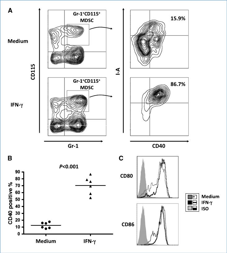
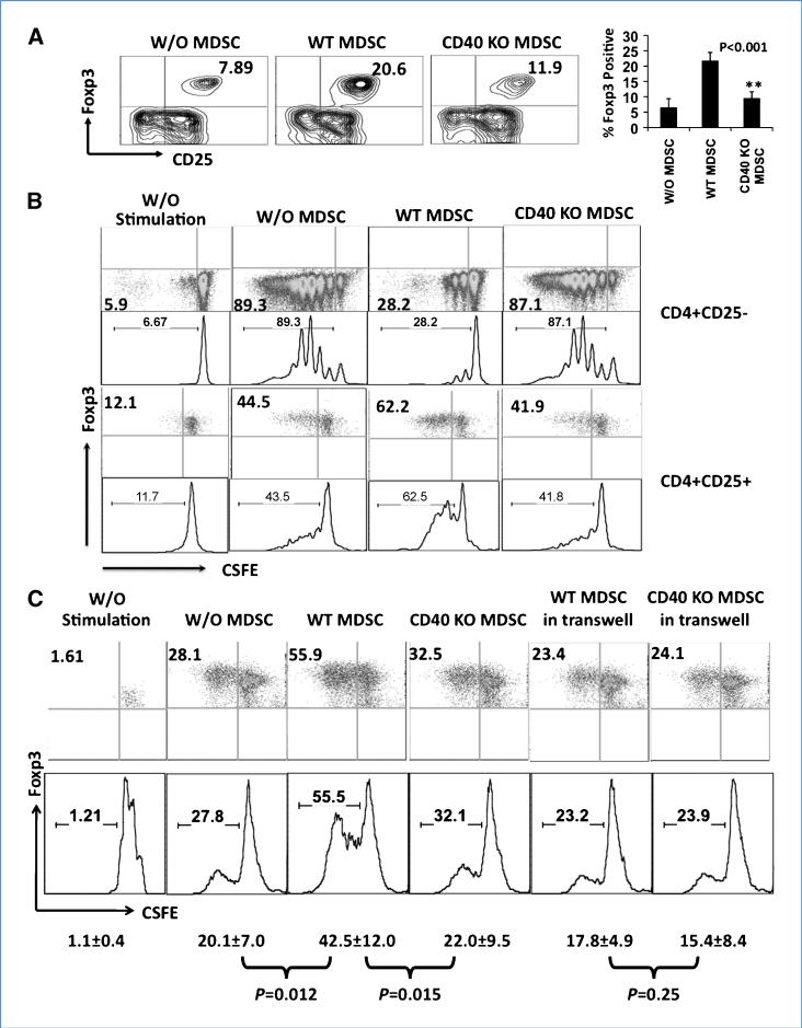
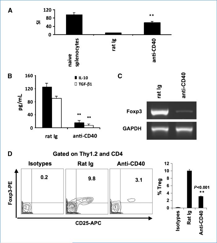
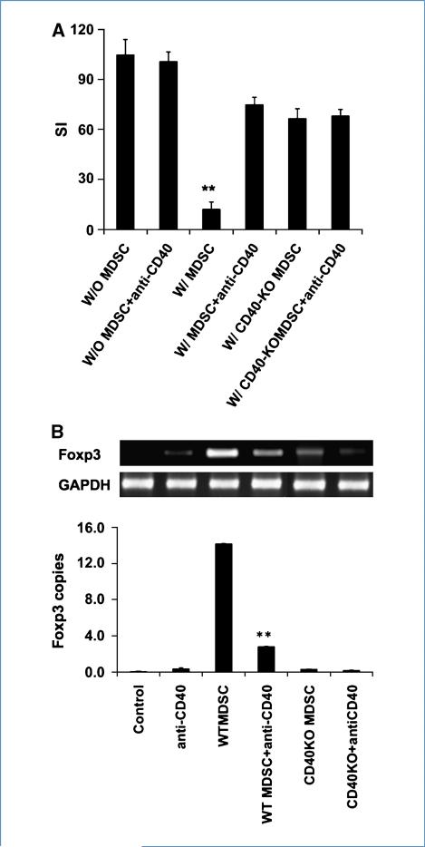
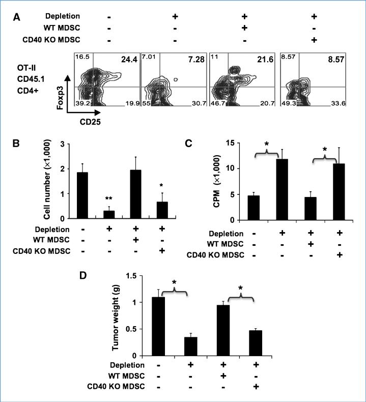
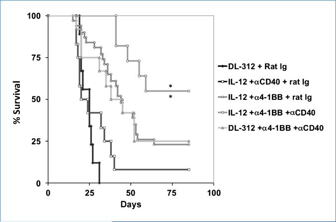
Immune tolerance to tumors is often associated with accumulation of myeloid-derived suppressor cells (MDSC) and an increase in the number of T-regulatory cells (Treg). In tumor-bearing mice, MDSCs can themselves facilitate the generation of tumor-specific Tregs. In this study, we demonstrate that expression of the immune stimulatory receptor CD40 on MDSCs is required to induce T-cell tolerance and Treg accumulation. In an immune reconstitution model, adoptive transfer of Gr-1+CD115+ monocytic MDSCs derived from CD40-deficient mice failed to recapitulate the ability of wild-type MDSCs to induce tolerance and Treg development in vivo. Agonistic anti-CD40 antibodies phenocopied the effect of CD40 deficiency and also improved the therapeutic efficacy of IL-12 and 4-1BB immunotherapy in the treatment of advanced tumors. Our findings suggest that CD40 is essential not only for MDSC-mediated immune suppression but also for tumor-specific Treg expansion. Blockade of CD40-CD40L interaction between MDSC and Treg may provide a new strategy to ablate tumoral immune suppression and thereby heighten responses to immunotherapy.


Figures

Figure 1
Figure 1
Expression of costimulatory molecules and MHC II by MDSC upon IFN-γ stimulation. Bone marrow Percoll fraction 2 cells, which contain MDSC, were cultured in the presence or absence of IFN-γ (100 ng/mL). Twenty-four hours later, cells were stained with fluorochrome-conjugated anti-Gr-1, anti-CD115, anti-CD40, anti-CD80, anti-CD86, and anti-I-Ab or isotype control. A, induction of CD40 on MDSC by IFN-γ. Flow cytometric data obtained from one representative experiment are presented as dot plots. B, significant induction of CD40 by IFN-γ. The results obtained from six tumor-bearing mice are presented. C, constitutive expression of CD80 and CD86 on MDSC.
Figure 2
Figure 2
Requirement of CD40 for MDSC-mediated T-cell suppression and Treg expansion in vitro. A, Treg induction by WT versus CD40 KO MDSCs. Purified CD4+ T cells were cocultured with WT or CD40 KO MDSCs at 4:1 ratio for 5 d followed by flow cytometry to assess the presence of CD4+CD25+Foxp3+ Tregs. Data are gated on CD4+ populations (n =6). B, expansion of CD4+CD25+Foxp3+ Tregs by WT, but not CD40 KO, MDSCs. CFSE-labeled, purified CD4+CD25 T cells and CD4+CD25+ Tregs from naïve mice were cocultured with WT or CD40 KO MDSCs at 4:1 ratio in the presence of anti-CD3/anti-CD28 for 3 d. Proliferation was assessed by flow cytometry. Histograms of CD4+CD25 population were gated on CD4+ cells, whereas those of CD4+CD25+ population were gated on Foxp3+ cells. Data from one representative of three reproducible experiments. C, CFSE-labeled, purified CD4+CD25+ Tregs from naïve OT-II mice were cocultured with WT or CD40 KO MDSCs at 4:1 (T cell to MDSC) ratio in the presence of irradiated OVA-EL4 cell (3,000 rad) at 10:1 ratio in the presence of IL-2 for 4 d. In the transwell experiment, MDSCs and Treg were added in the upper and lower chambers, respectively. Proliferation (CFSE dilution) was assessed by flow cytometry. Data from one representative of three reproducible experiments. Mean ± SD of each group has presented in results (n = 6).
Figure 3
Figure 3
Reversal of MDSC mediated immune suppression by anti-CD40. HA-TCR tumor (HA)–specific Thy-1.2 T cells were sorted from recipient HA-MCA26 tumor-bearing mice. Three mice per group were used in each of three reproducible and independent experiments. A, proliferative response of sorted Thy-1.2 T cells. Data are expressed as stimulation index (SI) relative to the cpm of T-cell proliferation in the absence of peptide (rat immunoglobulin versus anti-CD40; P < 0.001). B, IL-10 and TGF-β secretion by Thy-1.2 T cells. The concentrations of IL-10 and TGF-β in the culture supernatants were measured by ELISA (**, P < 0.001). C, prevention of Treg development by anti-CD40 in vivo. Foxp3 and glyceraldehyde-3-phosphate dehydrogenase (GAPDH) gene expression was assessed by RT-PCR of total RNA derived from sorted T cells. D, intracellular staining of Foxp3 in recovered tumor (HA)–specific T cells.
Figure 4
Figure 4
CD40 expression by MDSC is required for MDSC mediated tumor specific T cells immune suppression in vivo. After 9 d of adoptive transfer, tumor (HA)–specific CD4 Thy-1.2+ T cells were sorted from recipient HA-MCA26 tumor-bearing mice (Thy1.1+). One of three reproducible experiments was presented. Three mice per group were used in each of three independent experiments. A, proliferative response of sorted tumor-specific T cells. Data are expressed as stimulation index (SI) relative to the cpm of T-cell proliferation in the absence of peptide (W/MDSC versus W/O MDSC; P < 0.001, and no significant difference in other group). B, reduction in Foxp3 expression by tumor (HA)–specific T cells recovered from mice that also received CD40-deficient MDSCs. Foxp3 gene expression was assessed by RT-PCR on total RNA prepared from sorted T cells (WT MDSC versus WT MDSC + anti-CD40; P < 0.001).
Figure 5
Figure 5
CD40 is essential for Treg expansion, immune suppression, and tumor promotion mediated by MDSCs. OVA-B16–bearing MaFIA mice (CD45.2) were left untreated or depleted of CD115+ cells followed by adoptive transfer of CD45.1 OT-II T cells and reconstitution of WT or CD40 KO MDSC. A, tumor (OVA)–specific CD4+CD25+Foxp3+ Tregs in the tumor. Tumor infiltrated leukocytes were isolated and stained with antibodies against CD45.1, CD4, CD25, and Foxp3 or isotype controls followed by flow cytometry. Contour plots gated on CD45.1+CD4+ population are presented. One of three reproducible experiments (n = 3–4 per group) was presented. B, the number of tumor (OVA)–specific Tregs in the tumor. The numbers of CD45.1+ OT-II Tregs in the tumor were calculated (second versus first column, P < 0.001; fourth versus third column, P = 0.004). C, proliferative response of tumor (OT-II)–specific T cells reisolated from the recipient MaFIA mice. CD45.1+ OT-II T cells were reisolated from the spleen of recipient mice and stimulated with OVA peptide (1 μg/mL) in the presence of irradiated naïve splenocytes for 3 d. [3H]Thymidine was added in the last 8 h of culture (second versus first column, P = 0.002; fourth versus third column, P = 0.009). D, tumor weight in various treatment groups. Tumors were resected from tumor-bearing mice and weighed (second versus first column, P = 0.011; fourth versus third column, P = 0.006). Data were combined from three reproducible experiments (n = 9).
Figure 6
Figure 6
Effect of the combination of anti-CD40 and Adv/mIL-12 plus anti-4-1BB mAb on antitumor immunity. Mice bearing large MCA26 tumors were randomly assigned to four treatment groups. The long-term survival rate of mice treated with Adv/mIL-12 + anti-4-1BB + anti-CD40 (n = 12) is significantly higher than that of mice treated with Adv/mIL-12 + anti-4-1BB + rat immunoglobulin (n = 20; **, P < 0.01, log-rank test) and that of mice treated with DL312 + anti-4-1BB + anti CD40 (n = 12) or Adv/mIL-12 + anti-CD40 + rat immunoglobulin, respectively (n = 12; **, P < 0.01, log-rank test). All of the mice in the DL312 + rat immunoglobulin treatment group (n = 10) died on or before day 30 after tumor implantation. The combined results from two separate and reproducible experiments are presented.

Similar articles

Cited by

References

    1. Gattinoni L, Powell DJ, Jr., Rosenberg SA, Restifo NP. Adoptive immunotherapy for cancer: building on success. Nat Rev Immunol. 2006;6:383–93. - PMC - PubMed
    1. Li Q, Pan PY, Gu P, Xu D, Chen SH. Role of immature myeloid Gr-1+ cells in the development of antitumor immunity. Cancer Res. 2004;64:1130–9. - PubMed
    1. Rodriguez PC, Hernandez CP, Quiceno D, et al. Arginase I in myeloid suppressor cells is induced by COX-2 in lung carcinoma. J Exp Med. 2005;202:931–9. - PMC - PubMed
    1. Serafini P, Meckel K, Kelso M, et al. Phosphodiesterase-5 inhibition augments endogenous antitumor immunity by reducing myeloid-derived suppressor cell function. J Exp Med. 2006;203:2691–702. - PMC - PubMed
    1. Curiel TJ. Tregs and rethinking cancer immunotherapy. J Clin Invest. 2007;117:1167–74. - PMC - PubMed

Publication types

Substances