. 2010 Feb 5;285(6):4224-4231.
doi: 10.1074/jbc.M109.081018. Epub 2009 Dec 8.

Celastrol inhibits Hsp90 chaperoning of steroid receptors by inducing fibrillization of the Co-chaperone p23

Affiliations

Celastrol inhibits Hsp90 chaperoning of steroid receptors by inducing fibrillization of the Co-chaperone p23

Ahmed Chadli et al. J Biol Chem. .

Abstract

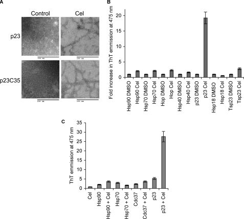
Hsp90 is an ATP-dependent molecular chaperone. The best characterized inhibitors of Hsp90 target its ATP binding pocket, causing nonselective degradation of Hsp90 client proteins. Here, we show that the small molecule celastrol inhibits the Hsp90 chaperoning machinery by inactivating the co-chaperone p23, resulting in a more selective destabilization of steroid receptors compared with kinase clients. Our in vitro and in vivo results demonstrate that celastrol disrupts p23 function by altering its three-dimensional structure, leading to rapid formation of amyloid-like fibrils. This study reveals a unique inhibition mechanism of p23 by a small molecule that could be exploited in the dissection of protein fibrillization processes as well as in the therapeutics of steroid receptor-dependent diseases.


Figures

FIGURE 1.
FIGURE 1.
Celastrol destabilizes the Hsp90 client PR in cells. A, HeLa PRB cells were treated for 4 h with various amounts of celastrol (Cel) or geldanamycin (GA). Cell lysates were prepared and blotted for the presence of Hsp90, Hsp70, and the Hsp90 clients. B, HeLa PRB cells were treated with 2.5 μm celastrol for various periods of time, and cell lysates were blotted for Hsp90 clients PRB and Akt. C, Hs578T cells were treated for 4 h with 5 μm celastrol, and cytosols were prepared and blotted for the presence of Hsp90, Hsp70, and the Hsp90 clients. AR, androgen receptor. D, HeLa PRB cells were treated with 2.5 μm celastrol for 4 h, and PRB stability was visualized by immunohistochemistry. DAPI, 4′,6-diamidino-2-phenylindole. E, effect of celastrol on the transcriptional activity of PR was assessed by measuring the production of the reporter gene CAT under control of two copies of the progesterone-response element in HeLa PRB cells. Cells were pretreated with 2.5 μm celastrol for 2 h prior to adding the progesterone agonist R5020 (125 nm) for an additional 10 h. Experiment was done twice, and each sample was done in triplicate.
FIGURE 2.
FIGURE 2.
Celastrol inhibits in vitro chaperoning of PR by affecting p23 function. A, PR hormone binding activity was reconstituted using purified chaperones in the presence of various amounts of celastrol (Cel, diamonds) or geldanamycin (GA, triangles). Binding of [3H]progesterone is expressed as a percentage of the DMSO-treated control. The experiment was done twice, and each sample was done in duplicate. B, celastrol has no effect on the ATPase activity of Hsp90. ATPase activity of Hsp90, stimulated by the addition of the activator Aha1, was measured in the presence of various concentrations of celastrol (Cel, diamonds) or geldanamycin (GA, triangles). Activity is expressed as the amount of [32P]ATP converted to [32P]ADP relative to the untreated control. Data are representative of two experiments. C, 20 μm purified chaperones were individually treated with 2-fold molar excess of celastrol prior to being used to reconstitute PR hormone binding activity. Excess celastrol was removed by extensive buffer exchange, and the treated chaperone was used with the other four untreated chaperones in reconstitution of PR. 5P represents reconstitution of PR with p23, Hsp90, Hsp70, Hop, and Hsp40 (Ydj1). The experiment was repeated three times. Each sample was done in triplicate. D, untreated p23 compensates for the celastrol-inactivated p23. Purified p23 was treated with five time molar excess of celastrol, and excess of celastrol was removed as in C. This celastrol-treated p23 was used in PR reconstitution reaction in the presence of indicated amounts of untreated p23. 4P stands for the four proteins Hsp90, Hsp70, Hop, and Hsp40 (Ydj1).
FIGURE 3.
FIGURE 3.
Celastrol destabilizes cellular Hsp90-p23 complexes. A, HeLa cells were treated with 3 μm celastrol, 17-allylamino-17-demethoxygeldanamycin (17AAG), or DMSO control for 18 h. Cell lysates were prepared, and Hsp90 immunoprecipitates (IP) were blotted for p23 or Hop. B, assessment of total Hsp90, Hsp70, or p23 in HeLa cell lysates shown in A. C, HeLa PRB and Hs578T cells were treated with control or siRNA against p23 for 96 h. Cytosols were blotted for p23, steroid receptors (PR, androgen receptor (AR), and GR), kinases (ChK1, RAF, CDK4, and ILK), Hsp70, and Hsp90.
FIGURE 4.
FIGURE 4.
Celastrol affects the structure of p23. A, 1H-15N heteronuclear single quantum coherence spectrum of 15N-labeled p23 recorded immediately after addition of a 5-fold molar excess of celastrol dissolved in DMSO is shown in red, and the corresponding spectrum after addition of the same amount of DMSO only is shown in black. Assigned resonances with weaker signals or disappeared signals following addition of celastrol are labeled. Prolonged treatment with celastrol led to the disappearance of all signals in the spectrum of p23 (data not shown). B, molecular surface representation of p23 (residues 1–119). The side chains of residues labeled in A are shown in red. The p23 area involved in binding Hsp90 is circled. C, 25 μm purified p23 was treated with 5-fold molar excess of celastrol or DMSO for 2 h at 37 °C and analyzed by size exclusion chromatography using a Superdex 200 column. Cel, celastrol.
FIGURE 5.
FIGURE 5.
Celastrol treatment causes p23 to form amyloid-like fibrils. A, 25 μm purified full-length p23 (p23WT) and its core domain p23C35 (residues 1–119) were treated with 2-fold molar excess of celastrol (Cel) and analyzed by electron microscopy. B, chaperone proteins Hsp90, Hsp70, Hop, Hsp40 (Ydj1), and Hsp18 along with p23 were treated with 2-fold molar excess of celastrol and analyzed for enhancement of thioflavin-T (ThT) fluorescence emission upon treatment with celastrol. The experiments were done two times, and all samples were tested in duplicate. C, Cdc37 treated with celastrol does not bind thioflavin-T. 25 μm Hsp90, Hsp70, Cdc37, and p23 were treated with 100 μm celastrol or DMSO control and incubated for 1 h at 37 °C and then 2 h at room temperature. Thioflavin-T binding was monitored using 96-well plates and the TECAN microplate reader at the excitation wavelength of 450 nm following emission at 475 nm.
FIGURE 6.
FIGURE 6.
Celastrol induces oligomerization of p23 in HeLa cells. A, immunocytochemistry analysis of cells treated with DMSO, 5 μm geldanamycin (GA), or celastrol (Cel) using the monoclonal antibody (JJ3) against p23. Yellow arrowheads indicate p23 clusters that are absent in the DMSO controls or geldanamycin-treated cells. The nucleus is stained with 4′,6-diamidino-2-phenylindole (blue) Scale bars represent 20 μm. B, immunoelectron microscopy analysis of cells treated with DMSO, 5 μm geldanamycin, or celastrol using the monoclonal antibody (Ab) (JJ3) against p23 and a gold-labeled goat anti-mouse antibody. Compared with DMSO or geldanamycin treatment, celastrol causes clustering of p23 that might indicate its fibrillization. Scale bars represent 200 nm. C, celastrol causes the redistribution of p23 into the insoluble fraction of HeLa cells. HeLa cells were treated with 3 μm celastrol or DMSO for 4 h and were fractionated into soluble (S) and insoluble pellet (P) fractions. Proportional amounts of each fraction were loaded and analyzed by Western blot for p23 and Hsp70.
FIGURE 7.
FIGURE 7.
Celastrol induction of p23 fibrils in wild-type and mutant p23. A, ability of wild-type full-length p23 and its core domain p23C35 (residues 1–119) to chaperone PR was compared with that of p23 having the mutation C58S or the three mutations C40S, C58S, and C75S (Triple). 4P represents reconstitution of PR with Hsp90, Hsp70, Hop, and Hsp40 (Ydj1). Data represent three experiments in duplicate. Cel, celastrol. B, fibril formation upon celastrol treatment of p23 triple mutant (C40S, C58S, and C75S) was monitored by electron microscopy. C, ability of dihydrocelastrol to induce p23 fibrils was tested by thioflavin-T (ThT) binding using inhibitor-to-protein ratios of 2, 4, or 8 as indicated. Data represent three experiments. D, ability of dihydrocelastrol to induce p23 fibrils was monitored by electron microscopy.

Similar articles

Cited by

References

    1. Balch W. E., Morimoto R. I., Dillin A., Kelly J. W. (2008) Science 319, 916–919 - PubMed
    1. Latchman D. S. (2001) Cardiovasc. Res. 51, 637–646 - PubMed
    1. Whitesell L., Lindquist S. L. (2005) Nat. Rev. Cancer 5, 761–772 - PubMed
    1. Chiosis G., Vilenchik M., Kim J., Solit D. (2004) Drug Discov. Today 9, 881–888 - PubMed
    1. Tsutsumi S., Neckers L. (2007) Cancer Sci. 98, 1536–1539 - PMC - PubMed

Publication types

MeSH terms

LinkOut - more resources