. 2010 Feb 12;285(7):4278-90.
doi: 10.1074/jbc.M109.036731. Epub 2009 Dec 8.

Transforming growth factor beta1 inhibits cystic fibrosis transmembrane conductance regulator-dependent cAMP-stimulated alveolar epithelial fluid transport via a phosphatidylinositol 3-kinase-dependent mechanism

Affiliations

Transforming growth factor beta1 inhibits cystic fibrosis transmembrane conductance regulator-dependent cAMP-stimulated alveolar epithelial fluid transport via a phosphatidylinositol 3-kinase-dependent mechanism

Jérémie Roux et al. J Biol Chem. .

Abstract

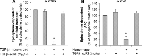
Exogenous or endogenous beta(2)-adrenergic receptor agonists enhance alveolar epithelial fluid transport via a cAMP-dependent mechanism that protects the lungs from alveolar flooding in acute lung injury. However, impaired alveolar fluid clearance is present in most of the patients with acute lung injury and is associated with increased mortality, although the mechanisms responsible for this inhibition of the alveolar epithelial fluid transport are not completely understood. Here, we found that transforming growth factor beta1 (TGF-beta1), a critical mediator of acute lung injury, inhibits beta(2)-adrenergic receptor agonist-stimulated vectorial fluid and Cl(-) transport across primary rat and human alveolar epithelial type II cell monolayers. This inhibition is due to a reduction in the cystic fibrosis transmembrane conductance regulator activity and biosynthesis mediated by a phosphatidylinositol 3-kinase (PI3K)-dependent heterologous desensitization and down-regulation of the beta(2)-adrenergic receptors. Consistent with these in vitro results, inhibition of the PI3K pathway or pretreatment with soluble chimeric TGF-beta type II receptor restored beta(2)-adrenergic receptor agonist-stimulated alveolar epithelial fluid transport in an in vivo model of acute lung injury induced by hemorrhagic shock in rats. The results demonstrate a novel role for TGF-beta1 in impairing the beta- adrenergic agonist-stimulated alveolar fluid clearance in acute lung injury, an effect that could be corrected by using PI3K inhibitors that are safe to use in humans.


Figures

FIGURE 1.
FIGURE 1.
TGF-β1 decreases β2AR agonist-stimulated alveolar fluid transport across primary rat ATII cell monolayers and in an in vivo model of hemorrhagic shock in rat. A, active TGF-β1 decreases epinephrine-dependent net fluid transport across polarized rat ATII cell monolayers. Rat ATII cell monolayers cultured at an air-liquid interface for 4 days were exposed to TGF-β1 (10 ng/ml) for 24 h; fluid absorption was calculated by the concentration of [125I]albumin (control 4.1% ± 0.9 of instilled, epinephrine (20 μm)-treated 7.3% ±1.1). The results are the means ± S.E. of at least nine monolayers from three experiments. *, p < 0.05 from cell monolayers exposed to TGF-β1 vehicle. B, TGF-β1 blockade restores the epinephrine-stimulated AFC in rats after hemorrhagic shock. The rats were pretreated intraperitoneally with TGF-β-soluble receptor (TGF-β-scRII; 2 mg/kg) or with its vehicle 30 min prior to hemorrhage or sham surgery and fluid resuscitation (AFC control 7.6% ± 1.4/30 min, epinephrine-treated 14.2% ± 2/30 min). The results are the means ± S.E. of six experiments for each animal group. *, p < 0.05 from control group.
FIGURE 2.
FIGURE 2.
TGF-β1 decreases β2AR agonist-stimulated CFTR-specific Cl transport across primary rat ATII cell monolayers. Short circuit current was measured in Ussing chambers in the presence of a Cl gradient after permeabilization of the basolateral membrane with nystatin (see “Experimental Procedures”). The total fraction of vectorial Cl transport through the apical membrane of rat ATII cells stimulated with epinephrine (20 μm) or terbutaline (20 μm) was inhibited by the CFTR inhibitor, CFTRinh-172 (10 μm). A, TGF-β1 decreases the epinephrine-stimulated CFTR-specific Cl absorption across the apical membrane of polarized rat ATII cells. A representative Ussing chamber recording (Isc) of polarized rat ATII cells treated or untreated with TGF-β1 is shown. B, TGF-β1 causes a time-dependent inhibition of the epinephrine-stimulated CFTR-specific Cl absorption across the apical membrane of polarized rat primary ATII cells. Pretreatment with a TGF-β soluble receptor II (TGF-β-scRII) specifically blocks TGF-β1 inhibition. C, TGF-β1 causes a dose-dependent inhibition of the epinephrine-stimulated CFTR-specific Cl absorption across the apical membrane of polarized rat ATII cells. D, TGF-β1 decreases the terbutaline-stimulated CFTR-specific Cl absorption across the apical membrane of polarized rat ATII cells. A representative Ussing chamber recording (Isc) of polarized rat ATII cells treated or untreated with TGF-β1 is shown. E, TGF-β1 causes a time-dependent inhibition of the terbutaline-stimulated CFTR-specific Cl absorption across the apical membrane of polarized rat primary ATII cells. Pretreatment with a TGF-β soluble receptor II (TGF-β-scRII) specifically blocks TGF-β1 inhibition. For all experiments, mean basal Isc was −11 ± 1.7 μA, mean epinephrine-treated Isc was −31 ± 2.1 μA, and mean terbutaline-treated Isc was −35 ± 1.8 μA in Cl gradient conditions; the results are the means ± S.E. of at least 12 monolayers from four experiments. *, p < 0.05 from monolayers exposed to TGF-β1 vehicle.
FIGURE 3.
FIGURE 3.
TGF-β1 decreases β2AR agonist-stimulated CFTR-specific Cl transport across primary human ATII cell monolayers. The short circuit current was measured in Ussing chambers in the presence of a Cl gradient after permeabilization of the basolateral membrane with nystatin (see “Experimental Procedures”). The total fraction of vectorial Cl transport through the apical membrane of human ATII cells stimulated with epinephrine (20 μm) was inhibited by the CFTR inhibitor, CFTRinh-172 (10 μm) (not shown). A, TGF-β1 causes a time-dependent inhibition of the epinephrine-stimulated CFTR-specific Cl absorption across the apical membrane of polarized human primary ATII cells. TGF-β-soluble receptor II (TGF-β-scRII) blocks specifically TGF-β1 inhibition. B, TGF-β1 causes a dose-dependent inhibition of the epinephrine-stimulated CFTR-specific Cl absorption across the apical membrane of polarized human ATII cells. For the two series of experiments, the mean basal Isc was −17 ± 2 μA, and the mean epinephrine-treated Isc was −34 ± 2.2 μA; the results are the means ± S.E. of at least 12 monolayers from four experiments. *, p < 0.05 from monolayers exposed to the TGF-β1 vehicle.
FIGURE 4.
FIGURE 4.
Short exposure (30 min) to TGF-β1 induces a heterologous desensitization of the β2AR and inhibits the β2AR-dependent activation of the cAMP/PKA pathway in primary rat ATII cell monolayers. A and B, short exposure (30 min) to TGF-β1 significantly decreases epinephrine (20 μm)-dependent cAMP and PKA activation in polarized rat primary ATII cells. cAMP content (control 4.1 ± 1.3 pmol/ml, epinephrine-treated 12.7 ± 2.4 pmol/ml) and PKA activity were measured in cell lysates by ELISA (control 1.9 ± 0.5 ng of active PKA/μg of protein, epinephrine-treated 4 ± 0.9). C, short exposure (30 min) to TGF-β1 does not affect the CPT-cAMP-dependent PKA activation at any concentration studied in polarized rat primary ATII cells. PKA activity was measured in cell lysates by ELISA (control 1.8 ± 0.4 ng active PKA/μg of protein). D, pretreatment with CPT-cAMP prevents TGF-β1-dependent decrease in β2AR agonist-stimulated CFTR-specific alveolar Cl transport across polarized rat primary ATII cell monolayers. Short circuit current was measured in Ussing chambers in the presence of a Cl gradient after permeabilization of the basolateral membrane with nystatin (see “Experimental Procedures”). The mean basal Isc was −10 ± 1.9 μA, and the mean epinephrine-treated Isc was −29 ± 2.6 μA; CPT-cAMP-treated Isc was −26 ± 1.7 μA in Cl gradient conditions. The results are the means ± S.E. of at least 12 monolayers from four experiments. *, p < 0.05 from monolayers exposed to TGF-β1 vehicle.
FIGURE 5.
FIGURE 5.
Short exposure (30 min) to TGF-β1 decreases β2AR agonist-stimulated Cl transport across primary rat ATII cell monolayers via a PI3K-dependent mechanism. A, short exposure (30 min) to TGF-β1 induces phosphorylation of Akt on serine 473 in polarized rat primary ATII cells. Phospho-Akt (Ser473) and total Akt protein expressions were measured by Western blot. B, short exposure (30 min) to TGF-β1 induces PI3Kα translocation to the plasma membrane in polarized rat primary ATII cells. Membrane PI3Kα and total PI3Kα protein expressions were measured by Western blot. C, pretreatment with PIK-90, a PI3K inhibitor, prevents the TGF-β1-mediated decrease in epinephrine-dependent cAMP in polarized rat primary ATII cells. cAMP content was measured in cell lysates by ELISA (control, 3.8 ± 1.2 pmol/ml; epinephrine-treated 11.9 ± 2.1, pmol/ml). D, PI3K inhibition (PIK-90, a specific PI3K inhibitor) prevents TGF-β1-dependent decrease in β2AR agonist-stimulated CFTR-specific alveolar Cl transport across polarized rat primary ATII cell monolayers. Isc was measured in Ussing chambers. E, short exposure (30 min) to TGF-β1 does not affect β2AR density at the cell membrane measured by saturation binding experiments ([125I]ICYP) (control Bmax = 533.5 ± 19.08 fmol/mg). ATII cell membranes were incubated with increasing concentrations of ICYP for 90 min at 37 °C. Nonspecific binding was determined in the presence of ICI-118,551 (100 μm). The values are representative of three independent experiments each performed in duplicate. F, short exposure (5 min) to TGF-β1 induces GRK2 protein translocation to the plasma membrane in polarized rat primary ATII cells without exposure to a β2-adrenergic agonist. GRK2 membrane and total protein expressions were measured by Western blot. G, short exposure (5 min) to TGF-β1 induces phosphorylation of the β2AR on its serine residue in position 355 in polarized rat primary ATII cells. Total and phospho-β2AR (Ser355), protein expressions were measured by Western blot. H, GRK2 inhibitor prevents the TGF-β1 short exposure-mediated inhibition of the epinephrine-stimulated CFTR-specific Cl absorption across the apical membrane of polarized rat primary ATII cells. Isc was measured in Ussing chambers. I, pretreatment with IBMX, a phosphodiesterase inhibitor (0.5 mm), prevents the TGF-β1 short exposure-induced decrease of the epinephrine-stimulated CFTR-specific Cl absorption across the apical membrane of polarized rat primary ATII cells. Short circuit current was measured in Ussing chambers in the presence of a Cl gradient after permeabilization of the basolateral membrane with nystatin (see “Experimental Procedures”). The mean basal Isc was −9 ± 2.2 μA, and the mean epinephrine-treated Isc was −29 ± 2.3 μA, in Cl gradient conditions. For all experiments, the results are the means ± S.E. of at least 12 monolayers from four experiments. *, p < 0.05 from monolayers exposed to TGF-β1 vehicle. For Western blot experiments, one representative experiment is shown, and three additional experiments gave comparable results; densitometry analysis results are the means ± S.E. of four experiments. *, p < 0.05 from monolayers exposed to TGF-β1 vehicle.
FIGURE 6.
FIGURE 6.
Prolonged exposure (6 h) to TGF-β1 inhibits the β2AR-dependent activation of the PKA and CFTR promoter activity, gene expression, and function via a PI3K-mediated down-regulation of the β2-adrenergic receptor at the cell membrane. A, prolonged exposure (6 h) to TGF-β1 decreases β2AR cell membrane density, measured by saturation binding experiments ([125I]ICYP). PI3K inhibition (PIK-90, a specific PI3K inhibitor) prevents TGF-β1-dependent decrease in β2AR plasma membrane density (control Bmax = 512.3 ± 17.28 fmol/mg; KD = 25.37 ± 3.087 pm; TGF-β1-treated Bmax = 305.9 ± 17.6 fmol/mg, KD = 25.23 ± 5.249 pm). ATII cell membranes were incubated with increasing concentrations of ICYP for 90 min at 37 °C. Nonspecific binding was determined in the presence of ICI-118,551 (100 μm). The values are representative of three independent experiments, each performed in duplicate. B, prolonged exposure (6 h) to TGF-β1 inhibits the epinephrine-dependent PKA activation in polarized rat primary ATII cells. PKA activity was measured in cell lysates by ELISA (control 2.1 ± 0.6 ng of active PKA/μg of protein, epinephrine-treated 4.7 ± 1.1). C, prolonged exposure (6 h) to TGF-β1 does not affect the CPT-cAMP-dependent PKA activation in polarized rat primary ATII cells. PKA activity was measured in cell lysates by ELISA (control 2.3 ± 0.7 ng of active PKA/μg of protein). D, PI3K inhibition (PIK-90, a specific PI3K inhibitor) prevents the TGF-β1-induced decrease in epinephrine-stimulated CFTR-specific Cl absorption across the apical membrane of polarized rat primary ATII cells. Short circuit current was measured in Ussing chambers in the presence of a Cl gradient after permeabilization of the basolateral membrane with nystatin (see “Experimental Procedures”). The mean basal Isc was −10 ± 2.6 μA, and the mean epinephrine-treated Isc was −30 ± 2.9 μA, in Cl gradient conditions. E, prolonged exposure (6 h) to TGF-β1 inhibits the epinephrine-stimulated but not the CPT-cAMP-stimulated CFTR promoter activity in rat ATII cells. CFTR promoter activity was measured in rat ATII cells transiently transfected with a CFTR-promoter reporter vector (38), containing the luciferase gene subcloned downstream of the CFTR promoter (CFTR(wt)-luc). Control luciferase activity = 88.7 ± 4.9 (RLU/μg protein). F, prolonged exposure (6 h) to TGF-β1 decreases the epinephrine-stimulated but not the CPT-cAMP-stimulated CFTR mRNA expression in polarized rat primary ATII cells. PI3K inhibition (PIK-90, a specific PI3K inhibitor) also prevents the TGF-β1-mediated decrease in the epinephrine-stimulated CFTR mRNA expression. CFTR mRNA levels were measured by real time RT-PCR normalized with glyceraldehyde-3-phosphate dehydrogenase mRNA levels. G, prolonged exposures (6 and 24 h) to TGF-β1 decrease the epinephrine-dependent CFTR protein expression at the plasma membrane of polarized rat primary ATII cells. The results are the means ± S.E. of at least 12 monolayers from four experiments. *, p < 0.05 from monolayers exposed to TGF-β1 vehicle; **, p < 0.05 from monolayers exposed to TGF-β1. For Western blot experiments, one representative experiment is shown, three additional experiments gave comparable results; densitometry analysis results are the means ± S.E. of four experiments. *, p < 0.05 from monolayers exposed to TGF-β1 vehicle.
FIGURE 7.
FIGURE 7.
PI3K inhibition prevents the TGF-β1-mediated decrease of the β2AR agonist-stimulated alveolar fluid transport in a rat model of hemorrhagic shock. A, hemorrhagic shock induces phosphorylation of Akt on serine 473 in rat lungs. Phospho-Akt (Ser473) and total Akt protein expressions were measured by Western blot. One representative experiment is shown, and three additional experiments gave comparable results; densitometry analysis results are the means ± S.E. of four experiments. *, p < 0.05 from control group. B, a specific PI3K inhibitor, PIK-90, restores the epinephrine stimulated AFC in rats after hemorrhagic shock. The rats were pretreated intraperitoneally with PIK-90 (5 mg/kg) or with its vehicle 30 min prior to hemorrhage or sham surgery and fluid resuscitation (AFC: control, 6.9% ±1.5/30 min; epinephrine-treated, 15.2% ±1.7/30 min). The results are the means ± S.E. of six experiments for each animal group. *, p < 0.05 from control group.

Similar articles

Cited by

References

    1. Rubenfeld G. D., Caldwell E., Peabody E., Weaver J., Martin D. P., Neff M., Stern E. J., Hudson L. D. (2005) N. Engl. J. Med. 353, 1685–1693 - PubMed
    1. Ware L. B., Matthay M. A. (2000) N. Engl. J. Med. 342, 1334–1349 - PubMed
    1. Ware L. B., Matthay M. A. (2001) Am. J. Respir. Crit. Care Med. 163, 1376–1383 - PubMed
    1. Calfee C. S., Matthay M. A. (2007) Chest 131, 913–920 - PMC - PubMed
    1. Berthiaume Y. (1991) J. Appl. Physiol. 70, 2490–2497 - PubMed

Publication types

MeSH terms

Substances