. 2010 Feb 5;285(6):3600-3607.
doi: 10.1074/jbc.M109.083139. Epub 2009 Dec 8.

Modulation of the human glucuronosyltransferase UGT1A pathway by splice isoform polypeptides is mediated through protein-protein interactions

Affiliations

Modulation of the human glucuronosyltransferase UGT1A pathway by splice isoform polypeptides is mediated through protein-protein interactions

Judith Bellemare et al. J Biol Chem. .

Abstract

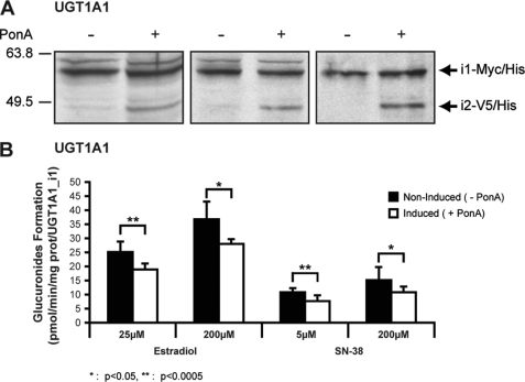
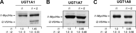
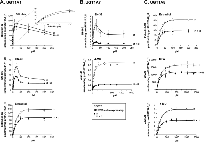
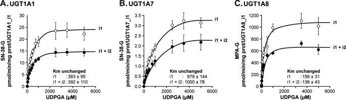
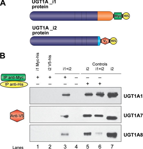
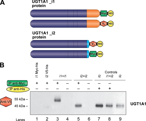
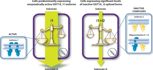
This study investigated the molecular mechanisms underlying the regulatory effect of the newly discovered 45-kDa enzymatically inactive UGT1A spliced polypeptides, named isoform i2, upon UGT1A-mediated glucuronidation. Initially, using an inducible system that mimics the relative abundance of isoforms 1 and 2 of UGT1A1 in human tissues, the rates of formation of glucuronides were significantly reduced. We then used a heterologous system constitutively expressing both isoforms i1 and i2 for an in-depth investigation of the presence of spliced i2 on glucuronidation kinetics. UGT1A1, UGT1A7, and UGT1A8 were selected as candidates for these studies. In all cases, co-expression of i1 and i2 in HEK293 cells leads to a significant reduction of the velocity of the glucuronidation reaction without affecting the affinity (K(m) (app)) for all substrates tested and the K(m) for the co-substrate, UDP-glucuronic acid. The data are consistent with a dominant-negative model of inhibition but do not sustain with an UGT1A_i2-mediated inhibition by competitive binding for substrate or the co-substrate. In contrast, the data from the co-immunoprecipitation experiments are indicative of the existence of a mixture homo-oligomeric (i1-i1 or i2-i2) and hetero-oligomeric (i1-i2) complexes in which the i2-i2 and i1-i2 subunits would be inactive. Thus, protein-protein interactions are likely responsible for the inhibition of active UGT1A_i1 by i2 spliced polypeptides. This new regulatory mechanism may alternatively modulate cellular response to endo/xeno stimulus.


Figures

FIGURE 1.
FIGURE 1.
Induction of UGT1A1 protein isoform 2 in UGT1A1_i1-overexpressing cell line. The biological response of i1-expressing cells to i2 expression was investigated by inducing i2 expression by treatment of the cells with ponasterone A (5 μm) at 24 h of induction. A, induction of UGT1A1 isoform 2 proteins following ponasterone A (PonA) treatment for 24 h was visualized by Western blot analysis. Three independent induction experiments are shown. B, induction of UGT1A1_i2 inhibits i1-mediated glucuronidation activity on both substrates tested. Glucuronide formation was quantified by liquid chromatography/mass spectrometric analysis. The data represent the means ± S.D. of three independent experiments performed in triplicate. *, p < 0.5; **, p < 0.005.
FIGURE 2.
FIGURE 2.
Western blot analyses of UGT1A1, UGT1A7, and UGT1A8 cell lines stably expressing i1 and co-expressing i1 and i2. The presence of a 58-kDa protein in the HEK293-UGT1A_i1-Myc/His lines and a 48-kDa protein in the HEK293-UGT1A_i1+i2-V5/His lines is confirmed. The relative abundance (OD units) of overexpressed proteins was quantified and is indicated.
FIGURE 3.
FIGURE 3.
Formation of glucuronides for typical UGT1A substrates by cell lines expressing UGT1A1 (A), UGT1A7 (B), and UGT1A8 (C) spliced isoforms. Glucuronide formation was normalized to UGT1A_i1 protein content assessed by Western blot. The data represent the means ± S.D. of two independent experiments performed at least in triplicate.
FIGURE 4.
FIGURE 4.
Formation of glucuronides for typical UGT1A substrates in the presence of varying concentrations of the co-substrate by cell lines expressing UGT1A1 (A), UGT1A7 (B), and UGT1A8 (C) spliced isoforms. Glucuronide formation was normalized to UGT1A_i1 protein content assessed by Western blot. The data represent the means ± S.D. of two independent experiments performed at least in triplicate. Similar Km values for the co-substrate UDPGA were calculated in the presence of i1+i2 compared with cells expressing only i1.
FIGURE 5.
FIGURE 5.
Direct interaction between UGT1A spliced forms as demonstrated by co-immunoprecipitation assays. A, UGT1A isoforms 1 were subcloned and tagged with Myc-His epitope, whereas isoforms 2 were tagged with V5-His epitope. B, co-immunoprecipitation assays for UGT1A1, UGT1A7, and UGT1A8. Lanes 1 and 2, negative controls; immunoprecipitation (IP) with anti-Myc (1 μg). Lane 1, UGT1A_i1-Myc/His; lane 2, UGT1A_i2-V5/His; lanes 3 and 4, UGT1A_i1-Myc/His + UGT1A_i2-V5/His; lanes 5 and 6, positive controls immunoprecipitations with anti-His (1 μg); lane 5, visualization of UGT1A_i2-V5/His; lane 6, UGT1A_i2-V5/His (5 μg) was loaded onto the gel as positive control for the Western blot.
FIGURE 6.
FIGURE 6.
Homo-oligomerization of UGT1A1 spliced forms as demonstrated by co-immunoprecipitation assays. A, UGT1A1_i1 tagged with V5-His epitope and 1A1_i2 tagged with Myc-His epitope constructs were used in these assays. B, co-immunoprecipitation assays for UGT1A1. Lanes 1 and 2, negative controls; immunoprecipitation (IP) with anti-Myc (1 μg). Lane 1, UGT1A1_i1-Myc/His; lane 2, UGT1A1_i2-V5/His; lanes 3 and 4, UGT1A1_i1-Myc/His+UGT1A1_i1-V5/His; lanes 5 and 6, UGT1A1_i2-Myc/His + UGT1A1_i2-V5/His; lanes 7 and 8, positive controls immunoprecipitations with anti-His (1 μg); lane 5, UGT1A1_i2-V5/His; lane 8, UGT1A1_i1-Myc/His_1A1_i2-V5/His; lane 9, UGT1A1_i2-V5/His (5 μg) was loaded onto the gel as positive control for the Western blot.
FIGURE 7.
FIGURE 7.
Schematic representation of the dominant-negative regulatory character of UGT1A_i2 spliced isoform, which is proposed to occur through the formation of inactive complexes with active isoforms 1. In this model, the relative abundance of active (i1-i1)/inactive (i1-i2 and i2-i2) complexes would be determinant of global transferase activity of the cell.

Similar articles

Cited by

References

    1. Stamm S. (2008) J. Biol. Chem. 283, 1223–1227 - PubMed
    1. Wang E. T., Sandberg R., Luo S., Khrebtukova I., Zhang L., Mayr C., Kingsmore S. F., Schroth G. P., Burge C. B. (2008) Nature 456, 470–476 - PMC - PubMed
    1. Girard H., Lévesque E., Bellemare J., Journault K., Caillier B., Guillemette C. (2007) Pharmacogenet. Genomics 17, 1077–1089 - PubMed
    1. Lévesque E., Girard H., Journault K., Lépine J., Guillemette C. (2007) Hepatology 45, 128–138 - PubMed
    1. Gong Q. H., Cho J. W., Huang T., Potter C., Gholami N., Basu N. K., Kubota S., Carvalho S., Pennington M. W., Owens I. S., Popescu N. C. (2001) Pharmacogenetics 11, 357–368 - PubMed

Publication types

MeSH terms

LinkOut - more resources