Subretinal fluid in acute posterior multifocal placoid pigment epitheliopathy
- PMID: 19996817
- PMCID: PMC3695592
- DOI: 10.1097/IAE.0b013e3181c596f8
Subretinal fluid in acute posterior multifocal placoid pigment epitheliopathy
Abstract
Purpose: The purpose of this study was to describe the clinical finding of macular subretinal fluid by optical coherence tomography in patients with acute posterior multifocal placoid pigment epitheliopathy.
Methods: Patients with acute posterior multifocal placoid pigment epitheliopathy were identified, and those with macular serous retinal detachment noted clinically and confirmed by optical coherence tomography are described.
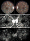
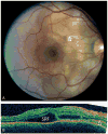
Results: Of 8 patients with acute posterior multifocal placoid pigment epitheliopathy evaluated by the uveitis service at the Illinois Eye and Ear Infirmary between 2003 and 2008, 4 eyes of 3 patients presented with macular subretinal fluid. Confirmatory optical coherence tomography was performed in two patients.
Conclusion: Acute posterior multifocal placoid pigment epitheliopathy may present clinically with macular subretinal fluid. This finding can be confirmed and monitored with optical coherence tomography.
Conflict of interest statement
The authors do not have any proprietary or financial interests with this study.
Figures


Similar articles
-
Spectral domain optical coherence tomography classification of acute posterior multifocal placoid pigment epitheliopathy.Retina. 2012 Jul;32(7):1403-10. doi: 10.1097/IAE.0b013e318234cafc. Retina. 2012. PMID: 22466468
-
[Subretinal fluid in acute posterior multifocal placoid pigment epitheliopathy: A case report].J Fr Ophtalmol. 2018 Sep;41(7):e303-e305. doi: 10.1016/j.jfo.2017.11.030. Epub 2018 Aug 31. J Fr Ophtalmol. 2018. PMID: 30173872 French. No abstract available.
-
OPTICAL COHERENCE TOMOGRAPHY ANGIOGRAPHY SHOWS INNER CHOROIDAL ISCHEMIA IN ACUTE POSTERIOR MULTIFOCAL PLACOID PIGMENT EPITHELIOPATHY.Retin Cases Brief Rep. 2017 Winter;11 Suppl 1:S136-S143. doi: 10.1097/ICB.0000000000000473. Retin Cases Brief Rep. 2017. PMID: 27759710
-
Spectral domain optical coherence tomography and autofluorescence in a case of acute posterior multifocal placoid pigment epitheliopathy mimicking Vogt-Koyanagi-Harada disease: case report and review of literature.Ocul Immunol Inflamm. 2011 Feb;19(1):42-7. doi: 10.3109/09273948.2010.521610. Epub 2010 Oct 31. Ocul Immunol Inflamm. 2011. PMID: 21034311 Review.
-
Progression of choroidal atrophy in acute posterior multifocal placoid pigment epitheliopathy.Ophthalmologica. 1998;212(1):66-72. doi: 10.1159/000027264. Ophthalmologica. 1998. PMID: 9438590 Review.
Cited by
-
Multifocal electroretinographic evaluation of macular function in acute posterior multifocal placoid pigment epitheliopathy.Doc Ophthalmol. 2013 Jun;126(3):253-8. doi: 10.1007/s10633-013-9378-x. Epub 2013 Mar 8. Doc Ophthalmol. 2013. PMID: 23471725
-
Atypical case of acute posterior multifocal placoid pigment epitheliopathy with intraretinal fluid.BMJ Case Rep. 2023 Oct 5;16(10):e255464. doi: 10.1136/bcr-2023-255464. BMJ Case Rep. 2023. PMID: 37798044
-
Acute posterior multifocal placoid pigment epitheliopathy following COVID-19 infection.Am J Ophthalmol Case Rep. 2023 Mar;29:101790. doi: 10.1016/j.ajoc.2022.101790. Epub 2022 Dec 29. Am J Ophthalmol Case Rep. 2023. PMID: 36597447 Free PMC article.
-
Mechanisms, Pathophysiology and Current Immunomodulatory/Immunosuppressive Therapy of Non-Infectious and/or Immune-Mediated Choroiditis.Pharmaceuticals (Basel). 2022 Mar 24;15(4):398. doi: 10.3390/ph15040398. Pharmaceuticals (Basel). 2022. PMID: 35455395 Free PMC article. Review.
-
A case of acute posterior multifocal placoid pigment epitheliopathy demonstrating vogt-koyanagi-harada disease-like optical coherence tomography findings in the acute stage.Case Rep Ophthalmol. 2013 Oct 11;4(3):172-9. doi: 10.1159/000356051. eCollection 2013. Case Rep Ophthalmol. 2013. PMID: 24403900 Free PMC article.
References
-
- Gass JD. Acute posterior multifocal placoid pigment epitheliopathy. Arch Ophthalmol. 1968;80:177–185. - PubMed
-
- Borruat FX, Piguet B, Herbort CP. Acute posterior multifocal placoid pigment epitheliopathy following mumps. Ocul Immunol Inflamm. 1998;6:189–193. - PubMed
-
- Azar P, Jr, Gohd RS, Waltman D, Gitter KA. Acute posterior multifocal placoid pigment epitheliopathy associated with an adenovirus type 5 infection. Am J Ophthalmol. 1975;80:1003–1005. - PubMed
-
- Bodine SR, Marino J, Camisa TJ, Salvate AJ. Multifocal choroiditis with evidence of Lyme disease. Ann Ophthalmol. 1992;24:169–173. - PubMed
-
- Fine HF, Kim E, Flynn TE, et al. Acute posterior multifocal placoid pigment epitheliopathy following varicella vaccine. Br J Ophthalmol. 2008 Aug 26; [Epub ahead of print] - PubMed
Publication types
MeSH terms
Substances
Grants and funding
LinkOut - more resources
Full Text Sources
Medical

