Review
. 2010 Jan;11(1):47-59.
doi: 10.1038/nrg2703. Epub 2009 Dec 8.

Origin and evolution of the adaptive immune system: genetic events and selective pressures

Affiliations
Review

Origin and evolution of the adaptive immune system: genetic events and selective pressures

Martin F Flajnik et al. Nat Rev Genet. 2010 Jan.

Abstract

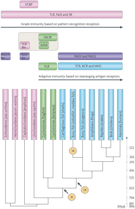
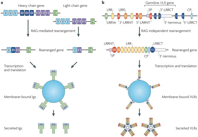
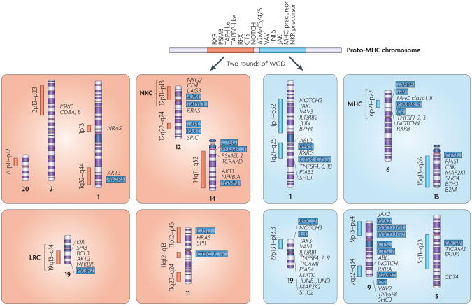
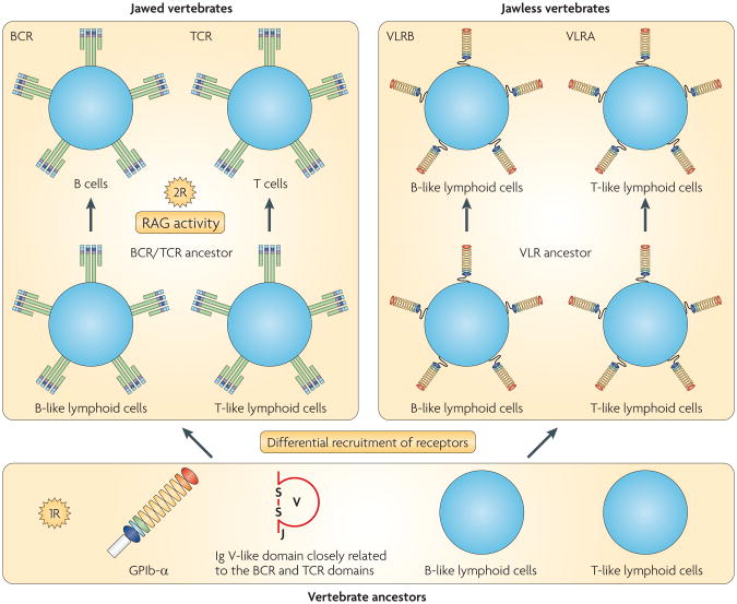
The adaptive immune system (AIS) in mammals, which is centred on lymphocytes bearing antigen receptors that are generated by somatic recombination, arose approximately 500 million years ago in jawed fish. This intricate defence system consists of many molecules, mechanisms and tissues that are not present in jawless vertebrates. Two macroevolutionary events are believed to have contributed to the genesis of the AIS: the emergence of the recombination-activating gene (RAG) transposon, and two rounds of whole-genome duplication. It has recently been discovered that a non-RAG-based AIS with similarities to the jawed vertebrate AIS - including two lymphoid cell lineages - arose in jawless fish by convergent evolution. We offer insights into the latest advances in this field and speculate on the selective pressures that led to the emergence and maintenance of the AIS.


Figures

Figure 1
Figure 1. Overview of the evolution of the immune system in deuterostomes
The stages in phylogeny at which the immune molecules referred to in this Review emerged. Molecules restricted to jawed and jawless vertebrates are indicated in blue and green, respectively. Molecules that emerged at the stage of invertebrates are in pink. Recombination-activating gene (RAG)-like genes (indicated in purple) are of viral or bacterial origin (from the transib transposon family) and are also present in the genomes of sea urchins and amphioxi. Agnathan paired receptors resembling antigen receptors (APAR) and novel immunoreceptor tyrosine-based activation motif-containing immunoglobulin superfamily receptor (NICIR, also known as T cell receptor (TCR)-like) are agnathan immunoglobulin superfamily (IgSF) molecules that are thought to be related to the precursors of TCRs and B cell receptors (BCRs). 1R and 2R indicate the two rounds of whole-genome duplication (WGD). Whether the 2R, the second round of WGD, occurred before or after the divergence of jawed and jawless vertebrates is controversial; the figure places it after the divergence according to the commonly held view (BOX 2). An ancestor of the majority of ray-finned fish is thought to have experienced an additional, lineage-specific WGD (designated as 3R) ∼320 million years ago,. The divergence time of animals (shown in Mya (million years ago)) is based on Blair and Hedges. MHC, major histocompatibility complex; NLR, Nod-like receptor; SR, scavenger receptor; TLR, Toll-like receptor; VCBP, V-region containing chitin-binding protein; VLR, variable lymphocyte receptor.
Figure 2
Figure 2. Antigen receptor proteins and genes in jawed vertebrates
a | The different B cell receptor (BCR) isotypes in mammals and their features in other vertebrates. Each oval represents an immunoglobulin superfamily (IgSF) domain consisting of 90-100 amino acids. Immunoglobulin D (IgD) is represented in two forms, mouse (left) and human (right); note that even in mammals the IgD structure is highly plastic. Major phylogenetic features are described in the text. b | BCR genes (which are defined by their heavy (H) chain) and T cell receptor (TCR) genes throughout phylogeny Recombination signal sequences are located at the 3′ end of all variable (V) loci, and the 5 end of joining (J) seqments and on both sides of diversity (D) segments. Note that in both the bony fish Ig H chain and vertebrate αδ TCRs, entire antigen receptor families are deleted upon rearrangement of the V segment to the downstream D and J segments (τ genes are deleted in bony fish and δ is deleted at the TCR locus). Note also that, despite its name, μ TCR in marsupials is related to δ TCR and not the IgM constant region. L chain, light chain; NAR, new antigen receptor.
Figure 3
Figure 3. Two distinct forms of adaptive immunity in vertebrates. a
a | Immunoglobulin (Ig) genes in jawed vertebrates. Ig genes generate repertoire diversity by recombining variable (V), diversity (D) and joining (J) gene segments with the help of the recombination-activating gene (RAG) recombinase. D gene segments occur only in the heavy chain genes. T cell receptors generate diversity in a similar manner (not shown). They occur only in a membrane-bound form. b | Variable lymphocyte receptors (VLRs) in jawless vertebrates. Multiple amino-terminal leucine-rich repeat (LRRNT)-, leucine-rich repeat (LRR)-, connecting peptide (CP)- and carboxy-terminal LRR (LRRCT)-encoding modules are located adjacent to the germline VLR gene. During the development of lymphoid cells, these modules are incorporated into the VLR gene. The rearranged VLR gene encodes a membrane-bound protein. VLRB has a secreted form and functions as an antibody, whereas VLRA apparently occurs only in a membrane-bound form. Whether membrane-bound VLRs occur as monomers or multimers is not known. LRR1 and LRRVe denote LRR modules located at the N- and C-termini, respectively. The organization of the VLR locus shows minor variations depending on loci and species. The figure is intended to emphasize the essential features of VLR assembly and does not accurately reproduce the organization of a specific VLR locus. SP, signal peptide.
Figure 4
Figure 4. The major histocompatibility complex paralogy group and the neurotrophin paralogy group in the human genome
Four sets of major histocompatibility complex (MHC) paralogons are located on chromoso mes 1, 6, 9 and 19 (ReFS 55,137,138). A number of smaller-sized MHC paralogons, which presumably originated from fragmentation and subsequent translocation of the major paralogons, have been identified. Among them, those located on 15q13-q26 and 5q11–q23 seem to have broken off from the paralogons on chromosomes 6 and 9, respectively. Four sets of major neurotrophin paralogons are located on chromosomes 1, 11, 12 and 19 (REF. 89).The MHC paralogy group (right) and neurotrophin paralogy group (left) are thought to have descended from neighbouring regions on a single ancestral chromosome. Paralogues that are distributed across the two paralogy groups are indicated by blue highlighted text. In addition to genes that share paralogues among the relevant paralogons, immunologically important genes, such as those encoding MHC class I, MHC class II, T cell receptors (TCRs), and immunoglobulins (Igs) are shown. A2M, α2-macroglobulin; B2M, β2-microglobulin; BCL, B cell leukaemia/lymphoma; CSK, cytoplasmic tyrosine kinase; CTS, cathepsin; ERAP, endoplasmic reticulum aminopeptidase; IL12RB, interleukin 12 receptor, β-subunit; JAK, Janus kinase; KIR, killer Ig-like receptor; LAG3, lymphocyte activation gene 3; LGMN, legumain; LRC, leukocyte receptor complex; MAP2K, mitogen-activated protein kinase kinase; MATK, megakaryocyte-associated tyrosine kinase; NFKBIA, nuclear factor-κB inhibitor; NKC, natural killer complex; NKR, NK receptor; NRAS, neuroblastoma RAS; PD, programmed cell death 1 ligand; PIAS, protein inhibitors of activated STAT; PIK3R, phosphatidylinositol 3-kinase, regulatory subunit; PSMB, proteasome subunits, β-type; PSME, proteasome activator subunits; RFX, regulatory factor X; RXR, retinoid X receptor; SPI1, spleen focus forming virus proviral integration oncogene; TAP, transporter associated with antigen processing; TAPBP, TAP-binding protein (also known as tapasin); TAPBPL, TAPBP-like; TAPL, TAP-like (also known as ABCB9); TICAM, TIR domain-containing adaptor molecule; TNFSF, tumour necrosis factor ligand superfamily; WGD, whole-genome duplication.
Figure 5
Figure 5. A hypothetical model for the origin of the two major forms of adaptive immune system
The occurrence of B- and T-like lymphoid cells in jawless vertebrates indicates that they were likely to be present in a common ancestor of all vertebrates. Similarly, the occurrence of a V-type immunoglobulin (Ig)-like domain with a canonical joining (J) segment in jawless vertebrates indicates that a common ancestor of all vertebrates had V-type Ig-like domains that could be converted to B cell receptor (BCR) or T cell receptor (TCR) Ig-like domains. Amphioxus, a basal chordate, has a glycoprotein Ib-α (GPIb-α)-like protein, which is the most likely precursor to the variable lymphocyte receptor (VLR). Therefore, a common ancestor of all vertebrates presumably had all of the key structural elements of the two radically different antigen receptors. Acquisition of recombination-activating gene (RAG) recombinase activities and the second round of whole-genome duplication (WGD; indicated as 2R) are thought to have had a crucial role in the emergence of the BCR–TCR–MHC-based adaptive immune system (AIS). Little is known about the genetic events that contributed to the emergence of the VLR-based AIS in jawless vertebrates. It is possible that the first round of WGD (indicated as 1R) contributed to the emergence of the two lineages of lymphoid cells; however, we cannot rule out the possibility that their origin predates the emergence of vertebrates.

Similar articles

Cited by

References

    1. Weigert MG, Cesari IM, Yonkovich SJ, Cohn M. Variability in the λ light chain sequences of mouse antibody. Nature. 1970;228:1045–1047. - PubMed
    1. Tonegawa S. Reiteration frequency of immunoglobulin light chain genes: further evidence for somatic ge neration of antibody diversity. Proc Natl Acad Sci USA. 1976;73:203–207. - PMC - PubMed
    1. Davis MM, Chien YH, Gascoigne NR, Hedrick SM. A murine T cell receptor gene complex: isolation, structure and rearrangement. Immunol Rev. 1984;81:235–258. - PubMed
    1. Oettinger MA, Schatz DG, Gorka C, Baltimore D. RAG-1 and RAG-2, adjacent genes that synergistically activate V(D)J recombination. Science. 1990;248:1517–1523. - PubMed
    1. Muramatsu M, et al. Class switch recombination and hypermutation require activation-induced cytidine deaminase (AID), a potential RNA editing enzyme. Cell. 2000;102:553–563. - PubMed

Publication types