. 2010 Feb;18(2):386-93.
doi: 10.1038/mt.2009.274. Epub 2009 Dec 8.

Complete genetic correction of ips cells from Duchenne muscular dystrophy

Affiliations

Complete genetic correction of ips cells from Duchenne muscular dystrophy

Yasuhiro Kazuki et al. Mol Ther. 2010 Feb.

Abstract

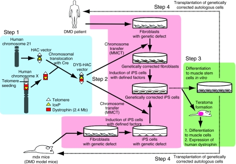
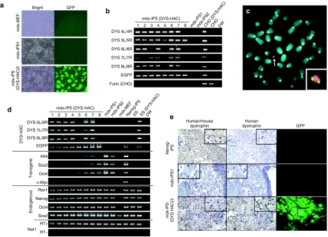
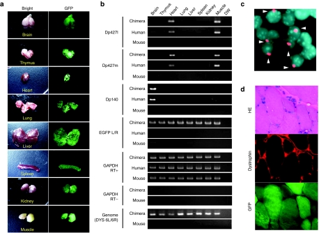
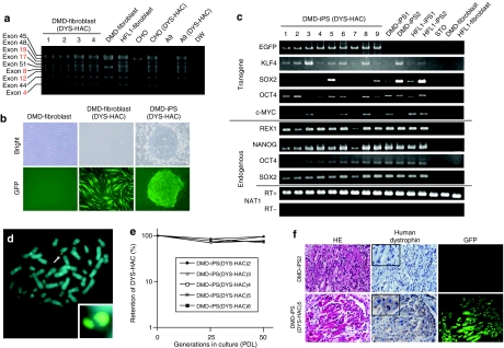
Human artificial chromosome (HAC) has several advantages as a gene therapy vector, including stable episomal maintenance that avoids insertional mutations and the ability to carry large gene inserts including the regulatory elements. Induced pluripotent stem (iPS) cells have great potential for gene therapy, as such cells can be generated from the individual's own tissues, and when reintroduced can contribute to the specialized function of any tissue. As a proof of concept, we show herein the complete correction of a genetic deficiency in iPS cells derived from Duchenne muscular dystrophy (DMD) model (mdx) mice and a human DMD patient using a HAC with a complete genomic dystrophin sequence (DYS-HAC). Deletion or mutation of dystrophin in iPS cells was corrected by transferring the DYS-HAC via microcell-mediated chromosome transfer (MMCT). DMD patient- and mdx-specific iPS cells with the DYS-HAC gave rise to differentiation of three germ layers in the teratoma, and human dystrophin expression was detected in muscle-like tissues. Furthermore, chimeric mice from mdx-iPS (DYS-HAC) cells were produced and DYS-HAC was detected in all tissues examined, with tissue-specific expression of dystrophin. Therefore, the combination of patient-specific iPS cells and HAC-containing defective genes represents a powerful tool for gene and cell therapies.


Figures

<b>Figure 1</b>
Figure 1
Schematic diagram of the HAC vector system for iPS cell–mediated gene therapy. Human dystrophin gene was cloned into a human chromosome 21–derived HAC vector using a combination of Cre-loxP-mediated chromosomal translocation and telomere-directed chromosomal truncation (Step 1, blue background). In Step 2 (pink background), mdx mice- or DMD patient–derived iPS cells were genetically restored by transfer of the DYS-HAC vector. The DYS-HAC was transferred to mdx-iPS cells directly. However, the DYS-HAC was transferred to DMD patient–derived fibroblasts via MMCT, as we failed to directly transfer the HAC into human iPS cells, then DMD-fibroblasts (DYS-HAC) were induced into iPS cells. Inability of MMCT into human iPS and embryonic stem cells is an unsolved issue. In Step 3 (green background), differentiation to muscle cells in vivo (teratoma formation) and expression of human dystrophin in muscle cells were confirmed. Step 4 represents the future and final goal of the transplantation of genetically corrected autologous cells by elegant differentiation and implantation technologies to come in a near future (dotted line). DMD, Duchenne muscular dystrophy; HAC, human artificial chromosome; iPS, induced pluripotent stem cells; Mb, megabase; MMCT, microcell-mediated chromosome transfer.
<b>Figure 2</b>
Figure 2
Characterization of mdx-iPS with DYS-HAC. (a) Morphology of mdx-MEF, mdx-iPS, and mdx-iPS (DYS-HAC) cells. Phase-contrast (left panel) and GFP-fluorescence (right panel) micrographs are shown. (b) Genomic PCR analyses for detecting DYS-HAC in mdx-iPS cells. (c) FISH analyses for mdx-iPS (DYS-HAC) cells. An arrow indicates the DYS-HAC and the inset shows an enlarged image of the DYS-HAC. (d) RT-PCR analyses of ES cell–marker genes, four exogenous transcription factors, and human dystrophin. EGFP and Nat1 were used as internal controls. Primers for DYS 6L/6R, 7L/7R, and 8L/8R detected the isoform of dystrophin expressed in ES and iPS cells. (e) Immunohistochemical analyses of dystrophin in muscle-like tissues of each teratoma. Immunodetection of mouse and human dystrophin (left panel), immunodetection of human-specific dystrophin (middle panel), and GFP micrography (right panel) are shown. The insets show enlarged images of immunohistochemistry. Nanog-iPS- and mdx-iPS-derived teratomas were used as positive and negative controls, respectively. CHO, Chinese hamster ovary; EGFP, enhanced green fluorescent protein; GFP, green fluorescent protein; HAC, human artificial chromosome; iPS, induced pluripotent stem cells; MEF, mouse embryonic fibroblast.
<b>Figure 3</b>
Figure 3
Expression analysis in chimeric mice with DYS-HAC. (a) Chimeric tissues derived from mdx-iPS (DYS-HAC). Bright (left panel) and fluorescence (right panel) micrographs are shown. (b) Representative genomic PCR and RT-PCR data for detection of DYS-HAC in each chimeric tissue. (c) FISH analysis of chimeric kidney derived from mdx-iPS (DYS-HAC) cells. Digoxigenin-labeled human COT-1 DNA (red) was used to detect DYS-HAC in tissues. Chromosomal DNA was counterstained with DAPI. Arrowheads show nuclei containing the DYS-HAC. (d) Immunohistochemical analyses of dystrophin in chimeric muscle. HE staining (top panel), immunodetection of dystrophin (middle panel), and GFP micrography (bottom panel) are shown. EGFP, enhanced green fluorescent protein; GFP, green fluorescent protein; HAC, human artificial chromosome; HE, hematoxylin and eosin; iPS, induced pluripotent stem cells.
<b>Figure 4</b>
Figure 4
Characterization of DMD-iPS with DYS-HAC. (a) Genomic multiplex PCR analyses for detecting the dystrophin genome on the DYS-HAC in DMD-fibroblast cells. Red lines show exons deleted in the DMD-fibroblast. (b) Morphology of DMD-fibroblasts, DMD-fibroblasts (DYS-HAC), and DMD-iPS (DYS-HAC) cells. Phase-contrast (top panel) and fluorescence (bottom panel) micrographs are shown. (c) RT-PCR analyses of embryonic stem cell–marker genes and four transcription factors. (d) FISH analyses for DMD-iPS (DYS-HAC) cells. An arrow indicates the DYS-HAC and the inset shows an enlarged image of the DYS-HAC. (e) Mitotic stability of the DYS-HAC in DMD-iPS (DYS-HAC) cells. (f) Immunohistochemical analyses of dystrophin in muscle-like tissues of each teratoma. HE staining (left panel), immunodetection of dystrophin (middle panel), and GFP micrography (right panel) are shown. The insets show enlarged images of immunohistochemistry. CHO, Chinese hamster ovary; DMD, Duchenne muscular dystrophy; GFP, green fluorescent protein; HAC, human artificial chromosome; HE, hematoxylin and eosin; iPS, induced pluripotent stem cells; PDL, population doubling.

Comment in

References

    1. Evans MJ., and , Kaufman MH. Establishment in culture of pluripotential cells from mouse embryos. Nature. 1981;292:154–156. - PubMed
    1. Thomson JA, Itskovitz-Eldor J, Shapiro SS, Waknitz MA, Swiergiel JJ, Marshall VS, et al. Embryonic stem cell lines derived from human blastocysts. Science. 1998;282:1145–1147. - PubMed
    1. Dennis C. Cloning: mining the secrets of the egg. Nature. 2006;439:652–655. - PubMed
    1. Hall VJ, Stojkovic P., and , Stojkovic M. Using therapeutic cloning to fight human disease: a conundrum or reality. Stem Cells. 2006;24:1628–1637. - PubMed
    1. Takahashi K., and , Yamanaka S. Induction of pluripotent stem cells from mouse embryonic and adult fibroblast cultures by defined factors. Cell. 2006;126:663–676. - PubMed

Publication types