. 2009 Dec;5(12):e1000757.
doi: 10.1371/journal.pgen.1000757. Epub 2009 Dec 4.

Homeobox transcription factors are required for conidiation and appressorium development in the rice blast fungus Magnaporthe oryzae

Affiliations

Homeobox transcription factors are required for conidiation and appressorium development in the rice blast fungus Magnaporthe oryzae

Seryun Kim et al. PLoS Genet. 2009 Dec.

Abstract

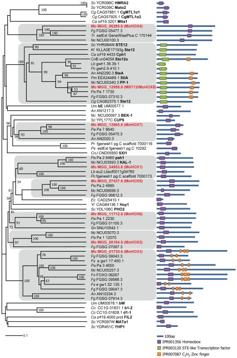
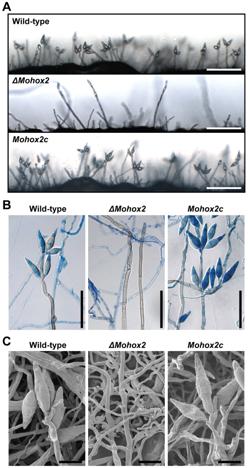
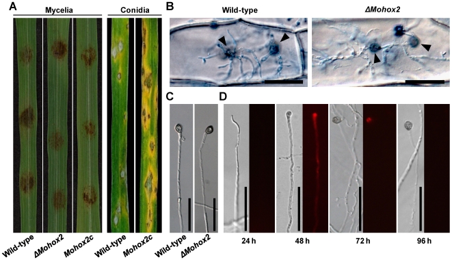
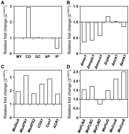
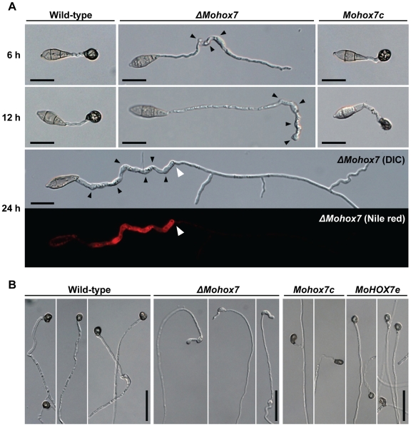
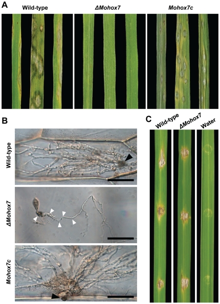
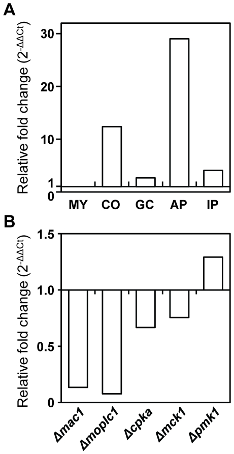
The appropriate development of conidia and appressoria is critical in the disease cycle of many fungal pathogens, including Magnaporthe oryzae. A total of eight genes (MoHOX1 to MoHOX8) encoding putative homeobox transcription factors (TFs) were identified from the M. oryzae genome. Knockout mutants for each MoHOX gene were obtained via homology-dependent gene replacement. Two mutants, DeltaMohox3 and DeltaMohox5, exhibited no difference to wild-type in growth, conidiation, conidium size, conidial germination, appressorium formation, and pathogenicity. However, the DeltaMohox1 showed a dramatic reduction in hyphal growth and increase in melanin pigmentation, compared to those in wild-type. DeltaMohox4 and DeltaMohox6 showed significantly reduced conidium size and hyphal growth, respectively. DeltaMohox8 formed normal appressoria, but failed in pathogenicity, probably due to defects in the development of penetration peg and invasive growth. It is most notable that asexual reproduction was completely abolished in DeltaMohox2, in which no conidia formed. DeltaMohox2 was still pathogenic through hypha-driven appressoria in a manner similar to that of the wild-type. However, DeltaMohox7 was unable to form appressoria either on conidial germ tubes, or at hyphal tips, being non-pathogenic. These factors indicate that M. oryzae is able to cause foliar disease via hyphal appressorium-mediated penetration, and MoHOX7 is mutually required to drive appressorium formation from hyphae and germ tubes. Transcriptional analyses suggest that the functioning of M. oryzae homeobox TFs is mediated through the regulation of gene expression and is affected by cAMP and Ca(2+) signaling and/or MAPK pathways. The divergent roles of this gene set may help reveal how the genome and regulatory pathways evolved within the rice blast pathogen and close relatives.


Conflict of interest statement

The authors have declared that no competing interests exist.

Figures

Figure 1
Figure 1. Phylogenetic analysis of putative homeobox transcription factors in fungi.
A neighbor-joining tree was constructed based on the amino acid sequences of representative fungal homeodomain TFs. The numbers at nodes represent the percentage of their occurrence in 10,000 bootstrap replicates; only nodes supported by 50 bootstraps or more are shown. The scale bar shows the number of amino acid differences per site. Subclades containing MoHOXs are shaded, in which MoHOXs and characterized genes from other fungi are shown in bold in red or black, respectively. Green and purple boxes in the domain architecture represent the homeodomains IPR001356 and IPR003120, respectively, and orange boxes are C2H2 zinc finger motifs (IPR007087). Abbreviations for fungal species followed by their GenBank accession numbers are as follows: Mo, Magnaporthe oryzae; Sc, Saccharomyces cerevisiae; Cg, Candida glabrata; Ca, Candida albicans; Fg, Fusarium graminearum; Tr, Trichoderma reesei; Nc, Neurospora crassa; Kl, Kluyveromyces lactis; CnB, Cryptococcus neoformans serotype D B3501-A; Lb, Laccaria bicolor; Pc, Phanerochaete chrysosporium; An, Aspergillus nidulans; Pm, Penicillium marneffeip; Pa, Podospora anserine; Um, Ustilago maydis; Pr, Phytophthora ramorum; Ps, Phytophthora sojae; CnJ, Cryptococcus neoformans serotype D JEC 21; Ec, Encephalitozoon cuniculi; Yl, Yarrowia lipolytica; Sn, Stagonospora nodorum; Fo, Fusarium oxysporum; Fs, Fusarium solani; Cc, Coprinus cinereus.
Figure 2
Figure 2. Effect of MoHOX2 deletion on conidiogenesis.
(A) Development of conidia on conidiophores. Light microscopic observation was performed on strains grown on V8 medium for 7 days. Bars = 100 µm. (B) Aerial structures stained with lactophenol aniline blue. Conidia and aerial hyphae stained blue, and conidiophores stained gray. Bars = 50 µm. (C) Scanned electron microscopic observation of aerial structures. Bars = 10 µm.
Figure 3
Figure 3. Effect of MoHOX2 deletion on appressorium development and pathogenicity.
(A) Assay for pathogenicity. Intact rice leaves were inoculated either by placement of agar plugs containing mycelia (6 mm in diameter) or by spraying conidial suspension (105 conidia/ml) of the indicated strains. (B) Assay for hypha-mediated penetration. Onion epidermis was inoculated with a patch of mycelia of the wild-type or ΔMohox2. Black arrowheads indicate sites of penetration via hyphal appressoria. Bars = 50 µm. (C) Appressorium development at hyphal tips. Mycelial blocks were placed on hydrophobic surfaces, and incubated for 72 h at 25°C. Bars = 50 µm. (D) Microscopic observation of temporal and spatial occurrence of lipid droplets during hypha-driven appressorium development. Mycelial blocks of the wild-type were incubated on hydrophobic surfaces, and stained with Nile red to visualize the formation of lipid droplets. Note that lipid droplets were not initially detected in hypha until 24 h, but became abundant in hypha with developing apprssorium 48 h after inoculation. Bars = 50 µm.
Figure 4
Figure 4. Transcriptional expression patterns of MoHOX2 and conidiogenesis-related genes.
(A) Expression of MoHOX2 during M. oryzae development. The tissues examined were submerged mycelia (MY), purified conidia (CO), germinated conidia (GC), appressoria (AP), and infected plants (IP). Measurements of MoHOX2 transcripts obtained by quantitative RT-PCR analysis were normalized to β-tubulin and expressed as relative values, with 1 corresponding to the MY. See Materials and Methods for details. (B) Expression of MoHOX2 in various deletion mutants. These include Δmac1, Δmoplc1, Δmocrz1, Δcpka1, Δmck1, and Δpmk1, and their genotypes were described in Table 1. The abundance of MoHOX2 transcripts in a mutant is expressed relative to a value of 1 in the wild-type KJ201. RNAs of the mutants were extracted from mycelia grown in CM liquid medium for 4 days. (C) Expression of conidiogenesis-related M. oryzae genes. The abundance of conidiogenesis-related transcripts in ΔMohox2 is expressed as a relative value, with 1 corresponding to MoHOX2 transcripts in the wild-type. (D) Expression of M. oryzae genes homologous to known conidiogenesis-related genes in other fungi. The abundance of conidiogenesis-related transcripts in ΔMohox2 is expressed as a relative value, with 1 corresponding to the level of MoHOX2 transcripts in the wild-type.
Figure 5
Figure 5. Effect of MoHOX7 deletion on appressorium formation.
(A) Microscopic observation of appressorium development on germ tubes. Appressorium formation was examined at 6, 12, and 24 h after incubation of the conidial suspension on hydrophobic cover slips. Black arrowheads in ΔMohox7 indicate the occurrence of swellings and hooks in a germ tube. Upon prolonged incubation of ΔMohox7 in the bottom panel, its germ tube initiated vegetative growth from a site indicated with a white arrowhead. The disappearance of lipid droplets after vegetative growth was revealed using the fluorescent dye Nile red. Bars = 20 µm. (B) Appressorium development on hyphae. Appressorium formation was induced by placing mycelia blocks on a hydrophobic plastic surface for 72 h. Bars = 50 µm.
Figure 6
Figure 6. Effect of MoHOX7 deletion on pathogenicity.
(A) Assay for pathogenicity. Conidial suspensions (105 conidia/ml) of the indicated strains were sprayed on 3-week-old intact rice leaves. Photographs were taken 7 days after inoculation. (B) Assay for penetration. 40 µl of conidial suspension (3×10⁴/ml) of the strains were inoculated on onion epidermal cells for 48 h. (C) Infiltration assay for growth inside plant cells. Conidial suspensions (5×10⁴ conidia/ml) of the indicated strains were infiltrated into rice leaves using a syringe. Water served as control. Photographs were taken 7 days after inoculation.
Figure 7
Figure 7. Expression patterns of MoHOX7.
(A) Expression of MoHOX7 during M. oryzae development. Abbreviations for the tissues used are shown in Figure 4A. The levels of MoHOX7 transcripts in a quantitative RT–PCR analysis were normalized to β-tubulin, and are expressed as relative values with 1 corresponding to the MY. (B) Expression of MoHOX7 in various deletion mutants. These include Δmac1, Δmoplc1, Δcpka1, Δmck1, and Δpmk1, and their genotypes were described in Table 1. The abundance of MoHOX7 transcripts in each mutant is expressed as a value relative to 1 in the wild-type KJ201.

References

    1. Ou SH. Rice Diseases. Kew, England: Commonwealth Mycological Institute; 1985. pp. 97–184.
    1. Ebbole DJ. Magnaporthe as a model for understanding host-pathogen interactions. Annu Rev Phytopathol. 2007;45:437–456. - PubMed
    1. Dean RA, Talbot NJ, Ebbole DJ, Farman ML, Mitchell TK, et al. The genome sequence of the rice blast fungus Magnaporthe grisea. Nature. 2005;434:980–986. - PubMed
    1. Valent B. Rice blast as a model system for plant pathology. Phytopathology. 1990;80:33–36.
    1. Howard RJ, Ferrari MA, Roach DH, Money NP. Penetration of hard substrates by a fungus employing enormous turgor pressures. Proc Natl Acad Sci U S A. 1991;88:11281–11284. - PMC - PubMed

Publication types

MeSH terms