

doi: 10.2174/1875043500902010008.
Three Dimensional OCT in the Engineering of Tissue Constructs: A Potentially Powerful Tool for Assessing Optimal Scaffold Structure
Affiliations
- PMID: 19997536
- PMCID: PMC2789573
- DOI: 10.2174/1875043500902010008
Item in Clipboard
Three Dimensional OCT in the Engineering of Tissue Constructs: A Potentially Powerful Tool for Assessing Optimal Scaffold Structure
Open Tissue Eng Regen Med J.
2009.
Abstract
Optical Coherence Tomography (OCT) provides detailed, real-time information on the structure and composition of constructs used in tissue engineering. The focus of this work is the OCT three-dimensional assessment of scaffolding architecture and distribution of cells on it. PLGA scaffolds were imaged in two and three-dimensions, both seeded and unseeded with cells. Then two types of scaffolds were reconstructed in three dimensions. Both scaffolding types were examined at three different seeding densities. The importance of three-dimensional assessments was evident, particularly with respect to porosity and identification of asymmetrical cell distribution.
Figures
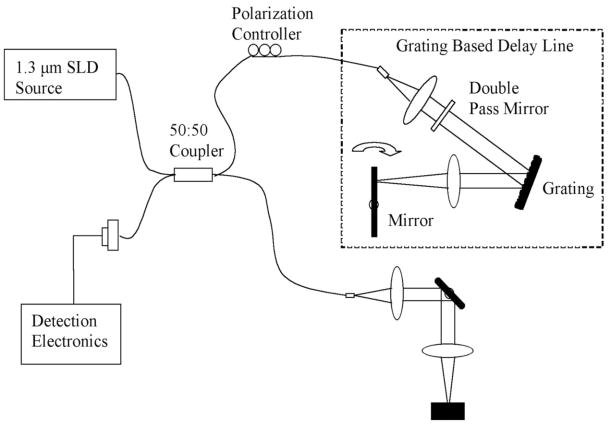
This image demonstrates a schematic of the OCT system used to generate images of the seeded and unseeded scaffolding. Imaging was performed at 10 frames per second that allowed rapid three-dimensional high-resolution reconstruction. The light source operates at a central frequency of 1300 nanometers that corresponds to an axial resolution of 10 microns measured from the point spread function off a mirror. The lateral resolution is approximately 25 microns. The scan rate is up to 3000 lines per second with a dynamic range of 100 decibels. The power on the sample was 10 milliwatts. An imaging catheter was used that was 0.019 inches in diameter with a focal line length of 2.0 millimeters. Recorded data was reconstructed in 3-D with Image J.
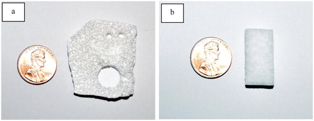
Photographs of unseeded PLGA (a) and Helistat scaffolding (b). The porosity can be noted.
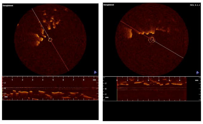
Figures 3a and b show two dimensional images of PLGA scaffolding unseeded and seeded, respectively. The scaffolds were seeded with human embryonic kidney cells (HEK-293). It can be seen that in two dimensions little detail is noted in the image and it is difficult, if not impossible, to assess both porosity and the degree of cell growth.
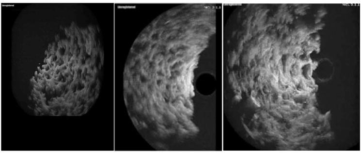
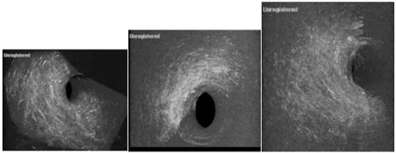
This figure shows unseeded (a), low density cell seeding (b), and high density cell seeding in PGLA scaffolding imaged in three dimensions. In 4a one rotational frame of the three dimensional scaffolding image is shown. We can see that pore size and distribution is well delineated compared to Fig. (3). In Fig. (3b) we see cell distribution in the scaffolding and the loss of porosity (arrow). This sample was seeded with 1,000,000 HEK-293 cells. In Fig. (3c) the scaffolding has been seeded with 2 million cells. In the periphery of the scaffolding there has been an almost complete loss in pores while the area most distant to the center still shows strong evidence of porosity, emphasizing asymmetrical distribution.
Three-dimensional imaging of the Helistat® scaffolding. In 5a a Helistat® scaffolding that consists of collagen type one, is shown in an unseeded sample. In image b, the scaffolding sample has been seeded with 1,000,000 cells (slightly higher due to the looseness of the collagen) and a reduction in the porosity is noted. However, it can be seen that the largest concentrations of cells occur on the outside of the Helistat® sponge with relatively little present in the interior. In image 5c, it is noted that where 2,000,000 cells are used to seed the graft the density has increased, however, the center of the graft again still remains relatively unseeded.
Similar articles
-
Three-dimensional, label-free cell viability measurements in tissue engineering scaffolds using optical coherence tomography.J Biomed Mater Res A. 2023 Aug;111(8):1279-1291. doi: 10.1002/jbm.a.37528. Epub 2023 Mar 14. J Biomed Mater Res A. 2023. PMID: 36916776
-
Optimal Seeding Densities for In Vitro Chondrogenesis of Two- and Three-Dimensional-Isolated and -Expanded Bone Marrow-Derived Mesenchymal Stromal Stem Cells Within a Porous Collagen Scaffold.Tissue Eng Part C Methods. 2016 Mar;22(3):208-20. doi: 10.1089/ten.TEC.2015.0365. Epub 2016 Jan 18. Tissue Eng Part C Methods. 2016. PMID: 26651081 Free PMC article.
-
Dynamic Assessment of the Endothelialization of Tissue-Engineered Blood Vessels Using an Optical Coherence Tomography Catheter-Based Fluorescence Imaging System.Tissue Eng Part C Methods. 2015 Jul;21(7):758-66. doi: 10.1089/ten.TEC.2014.0345. Epub 2015 Jan 30. Tissue Eng Part C Methods. 2015. PMID: 25539889 Free PMC article.
-
Imaging and characterization of bioengineered blood vessels within a bioreactor using free-space and catheter-based OCT.Lasers Surg Med. 2013 Aug;45(6):391-400. doi: 10.1002/lsm.22147. Epub 2013 Jun 5. Lasers Surg Med. 2013. PMID: 23740768
-
Short- and Long-term Evaluation of Bioresorbable Scaffolds by Optical Coherence Tomography.Interv Cardiol Clin. 2015 Jul;4(3):333-349. doi: 10.1016/j.iccl.2015.03.001. Epub 2015 May 29. Interv Cardiol Clin. 2015. PMID: 28581949 Review.
Cited by
-
Nondestructive Monitoring of Degradable Scaffold-Based Tissue-Engineered Blood Vessel Development Using Optical Coherence Tomography.J Vis Exp. 2018 Oct 3;(140):58040. doi: 10.3791/58040. J Vis Exp. 2018. PMID: 30346387 Free PMC article.
-
Imaging challenges in biomaterials and tissue engineering.Biomaterials. 2013 Sep;34(28):6615-30. doi: 10.1016/j.biomaterials.2013.05.033. Epub 2013 Jun 13. Biomaterials. 2013. PMID: 23768903 Free PMC article.
-
In-process monitoring of a tissue-engineered oral mucosa fabricated on a micropatterned collagen scaffold: use of optical coherence tomography for quality control.Heliyon. 2022 Nov 8;8(11):e11468. doi: 10.1016/j.heliyon.2022.e11468. eCollection 2022 Nov. Heliyon. 2022. PMID: 36406717 Free PMC article.
-
Taking a deep look: modern microscopy technologies to optimize the design and functionality of biocompatible scaffolds for tissue engineering in regenerative medicine.J R Soc Interface. 2013 Jul 17;10(86):20130263. doi: 10.1098/rsif.2013.0263. Print 2013 Sep 6. J R Soc Interface. 2013. PMID: 23864499 Free PMC article. Review.
-
Engineering next-generation oxygen-generating scaffolds to enhance bone regeneration.Trends Biotechnol. 2025 Mar;43(3):540-554. doi: 10.1016/j.tibtech.2024.09.006. Epub 2024 Sep 28. Trends Biotechnol. 2025. PMID: 39343620 Review.
References
-
- Lanza R, Langer R, Vacanti JP. Principles of Tissue Engineering. 2. Burlington; Massachusetts: 2000.
-
- Freed LE, Novakovic-Vunjak G. Tissue Engineering Bioreactors. In: Lanza RP, Langer R, Vacanti J, editors. Principles of tissue engineering. 2. Chap 23 Academic; San Diego:
-
- Frimberger D, Lin HK, Kropp BP. The use of tissue engineering and stem cells in bladder regeneration. Regen Med. 2006;1(4):425–35. Review. - PubMed
-
- Clark RA, Ghosh K, Tonnesen MG. Tissue engineering for cutaneous wounds. J Invest Dermatol. 2007;127(5):1018–29. Review. - PubMed
-
- Carrier RL, Rupnick MA, Langer R, Schoen FJ, Freed LE, Vunjak-Novakovic G. Effects of oxygen on engineered cardiac muscle. Biotech Bioeng. 2002;78(6):616–624. - PubMed
Grants and funding
LinkOut - more resources
Full Text Sources