

Spontaneous corneal perforation in a patient with lamellar ichthyosis and dry eye
- PMID: 19997563
- PMCID: PMC2788586
- DOI: 10.2147/opth.s8407
Spontaneous corneal perforation in a patient with lamellar ichthyosis and dry eye
Abstract
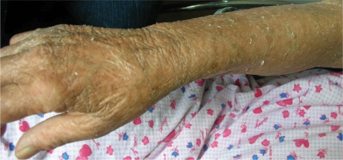
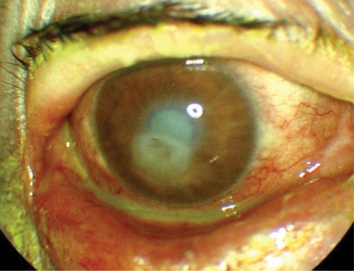
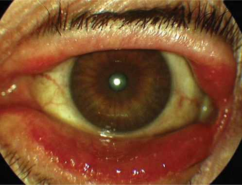
We report spontaneous corneal perforation in a patient with lamellar ichthyosis. The patient presented with complaints of pain, redness, diminished vision, and discharge in her right eye for 15 days. Visual acuities were light perception in the right and 20/400 in the left eye. Cicatricial ectropion in both lower eyelids and 2 mm perforation site in the center of the right cornea were observed. Lamellar ichthyosis was suspected because of scaling and excessive dryness of entire body skin and was confirmed by skin biopsy. Amniotic membrane transplantation and transient tarsorraphy was performed and systemic anti-ichthyosis therapy was started. The follow-up visits were not possible because of patient inconsistency. In patients with cicatricial ectropion secondary to ichthyosis, corneal health should be closely monitored because of the perforation risk.
Keywords: cornea; lamellar ichthyosis; spontaneous perforation.
Figures
Similar articles
-
Spontaneous bilateral corneal perforation in a patient with ichthyosis.Int Ophthalmol. 2014 Aug;34(4):919-21. doi: 10.1007/s10792-013-9866-8. Epub 2013 Oct 6. Int Ophthalmol. 2014. PMID: 24097116
-
Lamellar ichthyosis presenting as bilateral spontaneous corneal perforation.Nepal J Ophthalmol. 2013 Jan-Jun;5(1):117-9. doi: 10.3126/nepjoph.v5i1.7838. Nepal J Ophthalmol. 2013. PMID: 23584658
-
Case Report: Corneal Ulceration from Bilateral Ectropion Due to Congenital Ichthyosis.Optom Vis Sci. 2019 Sep;96(9):706-709. doi: 10.1097/OPX.0000000000001415. Optom Vis Sci. 2019. PMID: 31479026
-
[Cicatricial ectropion and ichthyosis. Apropos of 4 cases].J Fr Ophtalmol. 1992;15(5):349-56. J Fr Ophtalmol. 1992. PMID: 1430814 Review. French.
-
Diseases, conditions, and drugs associated with cicatricial ectropion.Arq Bras Oftalmol. 2019 May 20;82(4):345-353. doi: 10.5935/0004-2749.20190068. Arq Bras Oftalmol. 2019. PMID: 31116320 Review.
Cited by
-
Corneal Perforation as a Rare Ocular Manifestation in Lamellar Ichthyosis: Case Report and Literature Review.Indian Dermatol Online J. 2023 Oct 13;14(6):901-903. doi: 10.4103/idoj.idoj_679_22. eCollection 2023 Nov-Dec. Indian Dermatol Online J. 2023. PMID: 38099044 Free PMC article. No abstract available.
-
Spontaneous bilateral corneal perforation in a patient with ichthyosis.Int Ophthalmol. 2014 Aug;34(4):919-21. doi: 10.1007/s10792-013-9866-8. Epub 2013 Oct 6. Int Ophthalmol. 2014. PMID: 24097116
-
Biosynthesis of fatty aldehydes and alcohols in the eye and their role in meibogenesis.J Biol Chem. 2025 Jul;301(7):110330. doi: 10.1016/j.jbc.2025.110330. Epub 2025 Jun 3. J Biol Chem. 2025. PMID: 40473210 Free PMC article.
-
Cicatricial upper and lower eyelid ectropion in an ichthyosis patient. Surgical correction.J Dermatol Case Rep. 2011 Jun 6;5(2):27-9. doi: 10.3315/jdcr.2011.1068. J Dermatol Case Rep. 2011. PMID: 21894252 Free PMC article.
-
Ocular manifestation of Ichthyosis.Saudi J Ophthalmol. 2016 Jan-Mar;30(1):39-43. doi: 10.1016/j.sjopt.2015.12.004. Epub 2015 Dec 18. Saudi J Ophthalmol. 2016. PMID: 26949357 Free PMC article.
References
-
- Cruz AA, Menezes FA, Chaves R, Pinto Coelho R, Velasco EF, Kikuta H. Eyelid abnormalities in lamellar ichthyoses. Ophthalmology. 2000;107:1895–1898. - PubMed
-
- Esposito G, Tadini G, Paparo F, et al. Transglutaminase 1 deficiency and corneocyte collapse: an indication for targeted molecular screening in autosomal recessive congenital ichthyosis. Br J Dermatol. 2007;157:808–810. - PubMed
-
- Eltutar K, Karadede S, Atlihan F, Ozler S. Ectropion and bilateral corneal perforation in a case of congenital ichthyosiform erythroderma. Clinical and histopathologic review. Ophtalmologie. 1988;2:253–254. - PubMed
Publication types
LinkOut - more resources
Full Text Sources
