

JC virus mediates invasion and migration in colorectal metastasis
- PMID: 19997600
- PMCID: PMC2781631
- DOI: 10.1371/journal.pone.0008146
JC virus mediates invasion and migration in colorectal metastasis
Abstract
Introduction: JC Virus (JCV), a human polyomavirus, is frequently present in colorectal cancers (CRCs). JCV large T-Ag (T-Ag) expressed in approximately half of all CRC's, however, its functional role in CRC is poorly understood. We hypothesized that JCV T-Ag may mediate metastasis in CRC cells through increased migration and invasion.
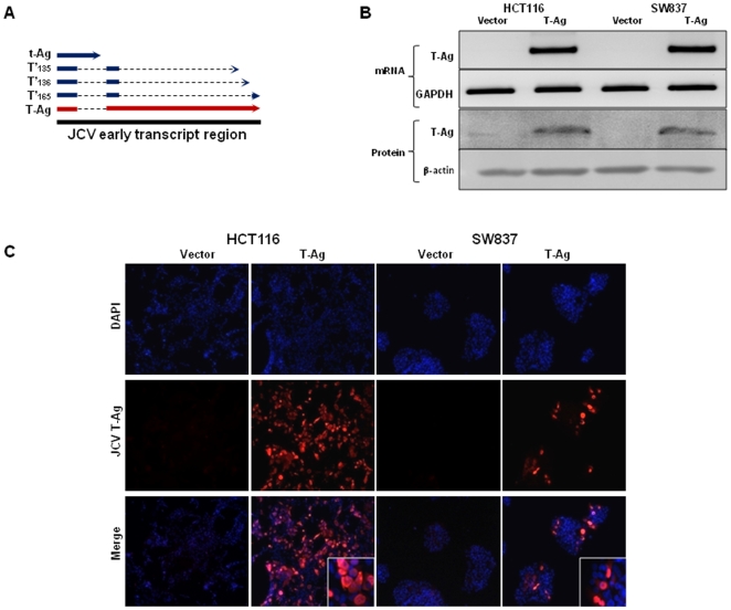
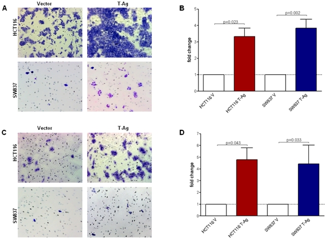
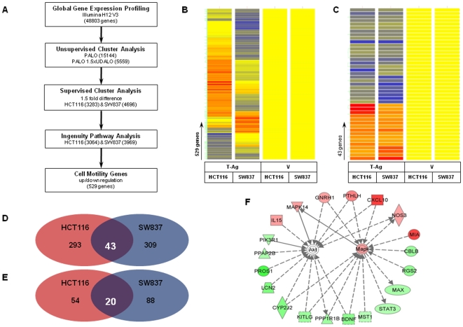
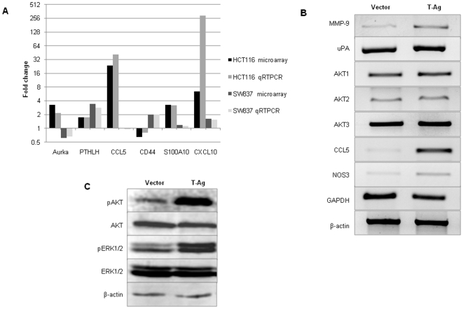
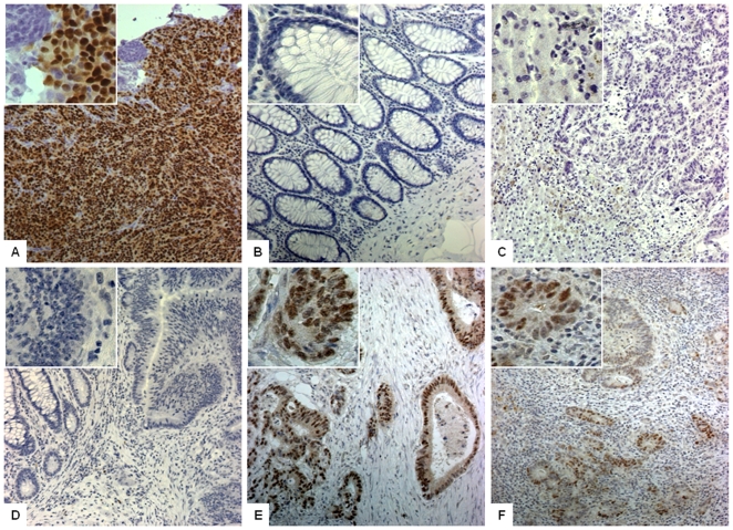
Material and methods: CRC cell lines (HCT116 and SW837) were stably transfected with JCV early transcript sequences cloned into pCR3 or empty vectors. Migration and invasion assays were performed using Boyden chambers. Global gene expression analysis was performed to identify genetic targets and pathways altered by T-Ag expression. Microarray results were validated by qRT-PCR, protein expression analyses and immunohistochemistry. Matching primary CRCs and liver metastases from 33 patients were analyzed for T-Ag expression by immunohistochemistry.
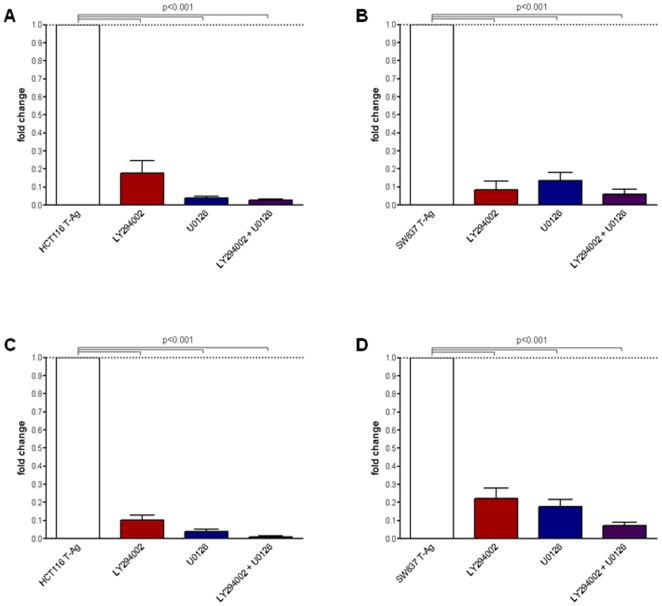
Results: T-Ag expressing cell lines showed 2 to 3-fold increase in migration and invasion compared to controls. JCV T-Ag expression resulted in differential expression of several genetic targets, including genes that mediate cell migration and invasion. Pathway analysis suggested a significant involvement of these genes with AKT and MAPK signaling. Treatment with selective PI3K/AKT and MAPK pathway inhibitors resulted in reduced migration and invasion. In support of our in-vitro results, immunohistochemical staining of the advanced stage tumors revealed frequent JCV T-Ag expression in metastatic primary tumors (92%) as well as in their matching liver metastasis (73%).
Conclusion: These data suggest that JCV T-Ag expression in CRC associates with a metastatic phenotype, which may partly be mediated through the AKT/MAPK signaling pathway. Frequent expression of JCV T-Ag in CRC liver metastasis provides further clues supporting a mechanistic role for JCV as a possible mediator of cellular motility and invasion in CRC.
Conflict of interest statement
Figures
Similar articles
-
MicroRNA miR-J1-5p as a potential biomarker for JC virus infection in the gastrointestinal tract.PLoS One. 2014 Jun 16;9(6):e100036. doi: 10.1371/journal.pone.0100036. eCollection 2014. PLoS One. 2014. PMID: 24932487 Free PMC article.
-
Association of JC virus T-antigen expression with the methylator phenotype in sporadic colorectal cancers.Gastroenterology. 2006 Jun;130(7):1950-61. doi: 10.1053/j.gastro.2006.02.061. Gastroenterology. 2006. PMID: 16762618
-
CHIP functions as an oncogene by promoting colorectal cancer metastasis via activation of MAPK and AKT signaling and suppression of E-cadherin.J Transl Med. 2018 Jun 19;16(1):169. doi: 10.1186/s12967-018-1540-5. J Transl Med. 2018. PMID: 29921293 Free PMC article.
-
JC Polyomavirus T-antigen protein expression and the risk of colorectal cancer: Systematic review and meta-analysis of case-control studies.PLoS One. 2023 Mar 31;18(3):e0283642. doi: 10.1371/journal.pone.0283642. eCollection 2023. PLoS One. 2023. PMID: 37000859 Free PMC article.
-
JC virus in the pathogenesis of colorectal cancer, an etiological agent or another component in a multistep process?Virol J. 2010 Feb 18;7:42. doi: 10.1186/1743-422X-7-42. Virol J. 2010. PMID: 20167111 Free PMC article. Review.
Cited by
-
Human Polyomavirus JCPyV and Its Role in Progressive Multifocal Leukoencephalopathy and Oncogenesis.Front Oncol. 2019 Aug 8;9:711. doi: 10.3389/fonc.2019.00711. eCollection 2019. Front Oncol. 2019. PMID: 31440465 Free PMC article. Review.
-
The Viral Janus: Viruses as Aetiological Agents and Treatment Options in Colorectal Cancer.Front Cell Infect Microbiol. 2021 Jan 7;10:601573. doi: 10.3389/fcimb.2020.601573. eCollection 2020. Front Cell Infect Microbiol. 2021. PMID: 33489934 Free PMC article.
-
Effect of the Large and Small T-Antigens of Human Polyomaviruses on Signaling Pathways.Int J Mol Sci. 2019 Aug 12;20(16):3914. doi: 10.3390/ijms20163914. Int J Mol Sci. 2019. PMID: 31408949 Free PMC article. Review.
-
MicroRNA miR-J1-5p as a potential biomarker for JC virus infection in the gastrointestinal tract.PLoS One. 2014 Jun 16;9(6):e100036. doi: 10.1371/journal.pone.0100036. eCollection 2014. PLoS One. 2014. PMID: 24932487 Free PMC article.
-
JC virus existence in Chinese gastrointestinal carcinomas.Oncol Lett. 2012 May;3(5):1073-1078. doi: 10.3892/ol.2012.627. Epub 2012 Feb 29. Oncol Lett. 2012. PMID: 22783394 Free PMC article.
References
-
- Jemal A, Siegel R, Ward E, Hao Y, Xu J, et al. Cancer statistics, 2009. CA Cancer J Clin 2009 - PubMed
-
- Steeg PS. Tumor metastasis: Mechanistic insights and clinical challenges. Nature Medicine. 2006;12:895–904. - PubMed
-
- Zur Hausen H. Oncogenic DNA viruses. Oncogene. 2001;20:7820–7823. - PubMed
-
- Knecht H, Berger C, Rothenberger S, Odermatt BF, Brousset P. The role of Epstein-Barr virus in neoplastic transformation. Oncology. 2001;60:289–302. - PubMed
Publication types
MeSH terms
Substances
Grants and funding
LinkOut - more resources
Full Text Sources
Medical
Molecular Biology Databases
