. 2010 Mar;67(5):781-96.
doi: 10.1007/s00018-009-0219-8. Epub 2009 Dec 9.

Identification of an emerin-beta-catenin complex in the heart important for intercalated disc architecture and beta-catenin localisation

Affiliations

Identification of an emerin-beta-catenin complex in the heart important for intercalated disc architecture and beta-catenin localisation

Matthew A Wheeler et al. Cell Mol Life Sci. 2010 Mar.

Abstract

How mutations in the protein emerin lead to the cardiomyopathy associated with X-linked Emery-Dreifuss muscular dystrophy (X-EDMD) is unclear. We identified emerin at the adherens junction of the intercalated disc, where it co-localised with the catenin family of proteins. Emerin bound to wild type beta-catenin both in vivo and in vitro. Mutating the GSK3beta phosphorylation sites on beta-catenin abolished this binding. Wild type but not mutant forms of emerin associated with X-EDMD were able to reduce beta-catenin protein levels. Cardiomyocytes from emerin-null mice hearts exhibited erroneous beta-catenin distribution and intercalated disc architecture. Treatment of wild type cardiomyocytes with phenylephrine, which inactivates GSK3beta, redistributed emerin and beta-catenin. Emerin was identified as a direct target of GSK3beta activity since exogenous expression of GSK3beta reduced emerin levels at the nuclear envelope. We propose that perturbation to or total loss of the emerin-beta-catenin complex compromises both intercalated disc function and beta-catenin signalling in cardiomyocytes.


Figures

Fig. 1
Fig. 1
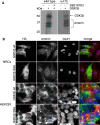
Characterisation of APS20 emerin antibody. a Lysates (20 μg) of C2C12s and NRCs, GST (5 μg) and GST-emerin 1-221 (5 μg) fusion proteins were resolved by SDS-PAGE and immunoblotted with AP8 and APS20. Blocking of APS20 was performed by pre-incubating the antibody with GST-emerin 114–183 fusion protein (40 μg) overnight. b Immunostaining of cryosectioned adult wild type and Emd −/y hearts (top two panels), NRCs (middle panel) and C2C12s (lower panel) with emerin antibodies. APS20 specificity was confirmed by pre-incubating antibody with emerin GST-114-183 fusion protein (APS20 + block). IDs are shown by arrows while the schematic depicts the region against which AP8 and APS20 were raised. Emd −/y hearts are stained with APS20 (red), β-catenin (green) and DAPI (blue). All images were captured on a Zeiss Axiovert inverted microscope, except the image labelled as ‘confocal’ which was captured on a Leica TCS SP5. Scale bars 10 μm
Fig. 2
Fig. 2
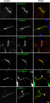
Emerin co-localises with specific junctional components of the intercalated disc. Immunostaining adult rat heart sections with APS20 (red) and for a range of ID (green) proteins illustrates emerin co-localises with α-catenin, β-catenin, plakoglobin and desmoplakin and not with αII-spectrin (arrow) and connexin 43 (arrow). Scale bar 10 μm
Fig. 3
Fig. 3
Emerin co-localises with the catenins at the adherens junction of intercalated discs (ID) and binds to β-catenin in vivo. a Electron micrographs of adult rat heart ID regions (arrow in i) in cryosections immunogold labelled for i APS20 (10 nm gold), ii APS20 pre-incubated with its antigen (10 nm gold), iii APS20 (10 nm gold) on Z-disc region of sarcomere, iv 10 and 5 nm gold-alone on ID, v APS20 (10 nm gold) and AP8 (5 nm gold), and vi APS20 (10 nm gold) on ID riser region (arrow; left hand panel) and desmosomes (arrow; right hand panel). Scale bars 0.1 μm. b Electron micrographs of adult rat ID regions in cryosections immunogold labelled for emerin and AJ proteins: i APS20 (10 nm gold) and γ-catenin (5 nm gold), ii APS20 (10 nm gold) and γ-catenin (5 nm gold), with arrowhead labelling the cytoplasmic plaque and arrow pointing to the terminal ends of the myofibrils at the AJ, iii APS20 (10 nm gold) and β-catenin (5 nm gold), iv APS20 (10 nm gold) and desmoplakin (5 nm gold). Scale bars i 0.1 μm, iiiii 0.05 μm. c AP8 (1:100) was added to NRC lysates to immunoprecipitate emerin and immunoblotted for β-catenin. I 5% total cell lysates input, B immunoprecipitated protein, A no primary antibody
Fig. 4
Fig. 4
Analysing NRCs treated with siRNA emerin (ad) and emerin null hearts for changes in morphology and catenin localisation (eh). NRCs were transfected with control siRNA, emerin siRNA or scrambled emerin siRNA for 72 h in medium containing PE. a Cells were fixed and immunostained with APS20 (red) to demonstrate reduced emerin at both the NE and the ID in the presence of emerin siRNA. Counterstaining for β-catenin (green) and DAPI (blue) reveals mislocalisation of β-catenin at the plasma membrane (arrowheads) in emerin siRNA treated cardiomyocytes compared with control cells. Scale bars 10 μm. b DIC images of the conditions shown in a. Arrows label the filopodial extensions. Scale bar 50 μm. c NRCs treated with control or emerin siRNA for 72 h were immunostained for emerin (red) and F-actin (green) and counterstained with DAPI (blue). d Immunoblot of control, scrambled and emerin siRNA treated NRCs demonstrates that reduced emerin expression does not alter total β-catenin levels. e Immunoblot of wild type and Emd −/y hearts shows that total β-catenin levels are unaffected by the loss of emerin. f Cryosections of 5-week-old wild type and Emd −/y mice hearts immunostained for either β- or γ-catenin. Insets represent higher magnification to illustrate increased convolution of the IDs in emerin-null mice. g Immunostaining of sections from 5-week-old wild type and Emd −/y mice hearts for β-catenin (green) and laminin (red). Insets represent higher magnification of IDs to illustrate β-catenin lateral staining. h The ratio of length to width of 80 cardiomyocytes from wild type and Emd −/y mice stained as for g were calculated using Image J. Data are shown from three individual mice as well as a mean value for each experimental group. **p < 0.001. Images were colleted on a Leica TCS SP5 confocal microscope for g only. ce Scale bars 10 μm
Fig. 5
Fig. 5
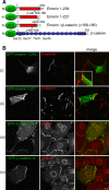
Emerin and β-catenin expression in NRCs. a Schematic diagram showing cDNA constructs used. Blue box represents the transmembrane, white-shadowed box the LEM domain, GFP-emerin full length 1-254, GFP-emerin 1-221, GFP-emerin Δ168-186 (β-catenin binding site [169-180] and surrounding residues deleted) and stabilised GFP-β-catenin (residues Ser33, Ser37, Thr41, Ser45 replaced with Ala). b NRCs in medium containing PE were transiently transfected with cDNA encoding i GFP-emerin 1-254, ii GFP-emerin 1-221, iii wild type GFP-β-catenin and iv stabilised GFP-β-catenin. Counterstaining for anti-β-catenin and anti-emerin is shown in red. Inset in panel i represents the captured image at a longer exposure and higher magnification of the ID region demonstrating low levels of GFP-emerin 1-254 at the ID. Scale bar 10 μm. Images were collected on a Leica TCS SP5 confocal microscope
Fig. 6
Fig. 6
Characterisation of the interaction between emerin and β-catenin. a Emerin binds to wild type but not stabilised β-catenin. Lysates of HEK293 transfected with GFP-emerin and GFP-β-catenin constructs were immunoprecipitated with AP8 and immunoblotted with anti-GFP antibody to visualise binding between emerin and β-catenin. GFP-β-cat st represents stabilised GFP-β-catenin construct. A representative blot from three independent experiments is shown. b Emerin regulates wild type but not stabilised β-catenin cellular levels through its interaction with β-catenin. Lysates of HEK293 cells were transfected with cDNA encoding GFP-emerin and GFP-β-catenin constructs and immunoblotted with anti-GFP. The mean band intensity of GFP-emerin and GFP-β-catenin constructs co-expressed was calculated as a percentage of their mean band intensities when expressed individually in the presence of GFP alone. c Emerin mutations disrupt its ability to regulate cellular β-catenin levels. HEK293 cells were co-transfected with wild type GFP-β-catenin and GFP-emerin constructs expressing X-EDMD mutations. Lysates were immunoblotted with anti-GFP antibody and mean band intensities for each GFP-construct calculated and tabulated as described for b above. d Emerin mutations disrupt the emerin–β-catenin affinity. Each GFP-emerin cDNA was expressed in HEK293 and normalised for protein levels and mixed with lysates containing an equal amount of GFP-β-catenin. Lysates were immunoprecipitated for β-catenin and immunoblotted for emerin. Binding is shown as a percentage of binding between wild type GFP-β-catenin and wild type GFP-emerin. For bd, results are shown as the mean of three independent experiments. *p < 0.05, **p < 0.01, ***p < 0.001
Fig. 7
Fig. 7
GSK3β phosphorylates emerin and regulates its intracellular location. a Emerin 1-221His6 (5 μg) and emerin 1-221His6 m175 (5 μg) were incubated in the absence (−) or presence (+) of 250U recombinant GSK3β in 1× reaction buffer for 30 min at 30°C. Several phospho-emerin bands are evident (M r 34–37 kDa) as is an auto-phosphorylated GSK3β (M r ~ 50 kDa). Inclusion of the GSK3β inhibitor SB216763 in the reaction completely abolished emerin phosphorylation. b Cultures of HEK293s and NRCs were transfected with cDNA encoding HA-tagged wild type GSK3β or HA-tagged GSK3β S9A. After 24 h cells were fixed. Immunostaining with anti-HA demonstrates exogenous expression of GSK3β whilst counterstaining with either APS20 (NRCs, red) or AP8 (HEK293s, green) demonstrates the loss of emerin from the NE in GSK3β expressing cells. Scale bars 10 μm
Fig. 8
Fig. 8
Stimulation of NRCs with PE relocates both β-catenin and emerin. ac NRCs pre-incubated in maintenance medium lacking PE for 24 h were then treated with 0.1% (v/v) DMSO, 100 μM PE or 100 μM PE + SB216763 for 30 min, 4 h and 24 h. Cardiomyocytes were fixed with 4% PFA and immunostained with APS20 (red), β-catenin (green) and DAPI (blue). Scale bar 10 μm. d Images collected from ac were imported into Image J, and the mean intensity of the nuclear region was calculated for both emerin and β-catenin staining in the presence of DMSO, PE or PE + SB216762. Images were taken from three independent experiments; n = 75. Images were captured on a Zeiss Axiovert inverted microscope

Similar articles

Cited by

References

    1. Emery AE. Emery-Dreifuss muscular dystrophy—a 40 year retrospective. Neuromuscul Disord. 2000;10:228–232. doi: 10.1016/S0960-8966(00)00105-X. - DOI - PubMed
    1. Bione S, Maestrini E, Rivella S, Mancini M, Regis S, Romeo G, Toniolo D. Identification of a novel X-linked gene responsible for Emery-Dreifuss muscular dystrophy. Nat Genet. 1994;8:323–327. doi: 10.1038/ng1294-323. - DOI - PubMed
    1. Bengtsson L, Wilson KL. Multiple and surprising new functions for emerin, a nuclear membrane protein. Curr Opin Cell Biol. 2004;16:73–79. doi: 10.1016/j.ceb.2003.11.012. - DOI - PubMed
    1. Ellis JA. Emery-Dreifuss muscular dystrophy at the nuclear envelope: 10 years on. Cell Mol Life Sci. 2006;63:2702–2709. doi: 10.1007/s00018-006-6247-8. - DOI - PMC - PubMed
    1. Holaska JM. Emerin and the nuclear lamina in muscle and cardiac disease. Circ Res. 2008;103:16–23. doi: 10.1161/CIRCRESAHA.108.172197. - DOI - PubMed

Publication types

MeSH terms

LinkOut - more resources