

Development of a scaffoldless three-dimensional engineered nerve using a nerve-fibroblast co-culture
- PMID: 19997868
- PMCID: PMC2864314
- DOI: 10.1007/s11626-009-9260-z
Development of a scaffoldless three-dimensional engineered nerve using a nerve-fibroblast co-culture
Abstract
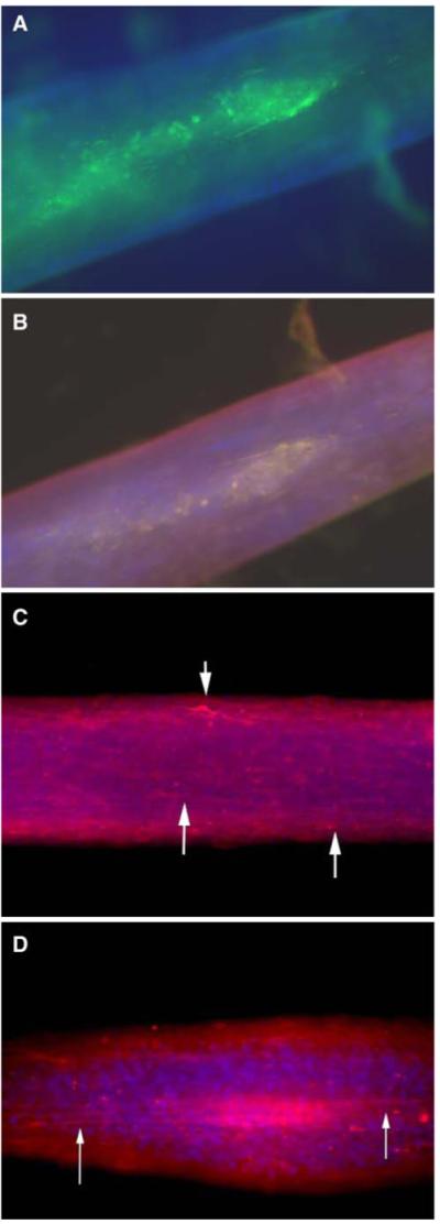
Nerve grafts are often required to replace tissue damaged by disease, surgery, or extensive trauma. Limitations such as graft availability, donor site morbidity, and immune rejection have led investigators to develop strategies to engineer nerve tissue. The goal of this study was to fabricate a scaffoldless three-dimensional (3D) nerve construct using a co-culture of fetal nerve cells with a fibroblast monolayer and allow the co-culture to remodel into a 3D construct with an external fibroblast layer and an internal core of interconnected neuronal cells. Primary fibroblasts were seeded on laminin-coated plates and allowed to form a confluent monolayer. Neural cells isolated from E-15 spinal cords were seeded on top of the fibroblast monolayer and allowed to form a networked monolayer across the monolayer of fibroblasts. Media shifts initiated contraction of the fibroblast monolayer and a remodeling of the co-culture into a 3D construct held statically in place by the two constraint pins. Immunohistochemistry using S100 (Schwann cell), beta3-tubulin, DAPI, and collagen I indicated an inner core of nerve cells surrounded by an external layer of fibroblasts. Conduction velocities of the 3D nerve and control (fibroblast-only) constructs were measured in vitro and compared to in vivo measures of neonatal sciatic nerve. The conduction velocities of the nerve constructs were comparable to 24-d-old neonatal nerve. The presence of Schwann cells and the ability to conduct neuronal signals in vitro suggest the scaffoldless 3D nerve constructs will be a viable option for nerve repair.
Figures


References
-
- Aszmann OC, et al. Bridging critical nerve defects through an acellular homograft seeded with autologous schwann cells obtained from a regeneration neuroma of the proximal stump. J Reconstr Microsurg. 2008;24(3):151–158. - PubMed
-
- Balgude AP, et al. Agarose gel stiffness determines rate of DRG neurite extension in 3D cultures. Biomaterials. 2001;22(10):1077–1084. - PubMed
-
- Bellamkonda R, et al. Hydrogel-based three-dimensional matrix for neural cells. J Biomed Mater Res. 1995;29(5):663–671. - PubMed
-
- Bryan DJ, et al. Enhanced peripheral nerve regeneration through a poled bioresorbable poly(lactic-co-glycolic acid) guidance channel. J Neural Eng. 2004;1(2):91–98. - PubMed
-
- Bunge MB, et al. Role of peripheral nerve extracellular matrix in Schwann cell function and in neurite regeneration. Dev. Neurosci. 1989;11(4–5):348–360. - PubMed
Publication types
MeSH terms
Substances
Grants and funding
LinkOut - more resources
Full Text Sources
Other Literature Sources
