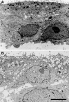
Cultures of human tracheal gland cells of mucous or serous phenotype
- PMID: 19998060
- PMCID: PMC2862963
- DOI: 10.1007/s11626-009-9262-x
Cultures of human tracheal gland cells of mucous or serous phenotype
Abstract
There are two main epithelial cell types in the secretory tubules of mammalian glands: serous and mucous. The former is believed to secrete predominantly water and antimicrobials, the latter mucins. Primary cultures of human airway gland epithelium have been available for almost 20 yr, but they are poorly differentiated and lack clear features of either serous or mucous cells. In this study, by varying growth supports and media, we have produced cultures from human airway glands that in terms of their ultrastructure and secretory products resemble either mucous or serous cells. Of four types of porous-bottomed insert tested, polycarbonate filters (Transwells) most strongly promoted the mucous phenotype. Coupled with the addition of epidermal growth factor (EGF), this growth support produced "mucous" cells that contained the large electron-lucent granules characteristic of native mucous cells, but lacked the small electron-dense granules characteristic of serous cells. Furthermore, they showed high levels of mucin secretion and low levels of release of lactoferrin and lysozyme (markers of native serous cells). By contrast, growth on polyethylene terephthalate filters (Cyclopore) in medium lacking EGF produced "serous" cells in which small electron-dense granules replaced the electron-lucent ones, and the cells had high levels of lactoferrin and lysozyme but low levels of mucins. Measurements of transepithelial resistance and short-circuit current showed that both "serous" and "mucous" cell cultures possessed tight junctions, had become polarized, and were actively secreting Cl.
Figures


Similar articles
-
Comparison of nonciliated tracheal epithelial cells in six mammalian species: ultrastructure and population densities.Exp Lung Res. 1983 Dec;5(4):281-94. doi: 10.3109/01902148309061521. Exp Lung Res. 1983. PMID: 6662075
-
Human tracheobronchial submucosal gland cells in culture.Am J Respir Cell Mol Biol. 1990 Jan;2(1):41-50. doi: 10.1165/ajrcmb/2.1.41. Am J Respir Cell Mol Biol. 1990. PMID: 2306368
-
Chloride secretion by cultures of pig tracheal gland cells.Am J Physiol Lung Cell Mol Physiol. 2012 May 15;302(10):L1098-106. doi: 10.1152/ajplung.00253.2011. Epub 2012 Feb 24. Am J Physiol Lung Cell Mol Physiol. 2012. PMID: 22367783 Free PMC article.
-
Isolation and culture of submucosal gland cells.Clin Chest Med. 1986 Jun;7(2):239-45. Clin Chest Med. 1986. PMID: 3522071 Review.
-
Regulation of secretion from serous and mucous cells in the trachea.Ciba Found Symp. 1984;109:4-19. doi: 10.1002/9780470720905.ch2. Ciba Found Symp. 1984. PMID: 6151486 Review.
Cited by
-
Cystic Fibrosis Human Organs-on-a-Chip.Micromachines (Basel). 2021 Jun 25;12(7):747. doi: 10.3390/mi12070747. Micromachines (Basel). 2021. PMID: 34202364 Free PMC article. Review.
-
CFTR and lung homeostasis.Am J Physiol Lung Cell Mol Physiol. 2014 Dec 15;307(12):L917-23. doi: 10.1152/ajplung.00326.2014. Epub 2014 Nov 7. Am J Physiol Lung Cell Mol Physiol. 2014. PMID: 25381027 Free PMC article. Review.
-
Development of glandular models from human nasal progenitor cells.Am J Respir Cell Mol Biol. 2015 May;52(5):535-42. doi: 10.1165/rcmb.2013-0259MA. Am J Respir Cell Mol Biol. 2015. PMID: 25412193 Free PMC article.
-
PAR-2-activated secretion by airway gland serous cells: role for CFTR and inhibition by Pseudomonas aeruginosa.Am J Physiol Lung Cell Mol Physiol. 2021 May 1;320(5):L845-L879. doi: 10.1152/ajplung.00411.2020. Epub 2021 Mar 3. Am J Physiol Lung Cell Mol Physiol. 2021. PMID: 33655758 Free PMC article.
-
Small-molecule activators of TMEM16A, a calcium-activated chloride channel, stimulate epithelial chloride secretion and intestinal contraction.FASEB J. 2011 Nov;25(11):4048-62. doi: 10.1096/fj.11-191627. Epub 2011 Aug 11. FASEB J. 2011. PMID: 21836025 Free PMC article.
References
-
- Cereijido M, Gonzalez-Mariscal L, Contreras G. Tight junction: barrier between higher organisms and environment. NIPS. 1989;4:72–76.

