. 2010 Jan;51(1):297-305.
doi: 10.1002/hep.23354.

Highly efficient generation of human hepatocyte-like cells from induced pluripotent stem cells

Affiliations

Highly efficient generation of human hepatocyte-like cells from induced pluripotent stem cells

Karim Si-Tayeb et al. Hepatology. 2010 Jan.

Erratum in

  • Hepatology. 2010 Mar;51(3):1094

Abstract

There exists a worldwide shortage of donor livers available for orthotropic liver transplantation and hepatocyte transplantation therapies. In addition to their therapeutic potential, primary human hepatocytes facilitate the study of molecular and genetic aspects of human hepatic disease and development and provide a platform for drug toxicity screens and identification of novel pharmaceuticals with potential to treat a wide array of metabolic diseases. The demand for human hepatocytes, therefore, heavily outweighs their availability. As an alternative to using donor livers as a source of primary hepatocytes, we explored the possibility of generating patient-specific human hepatocytes from induced pluripotent stem (iPS) cells.

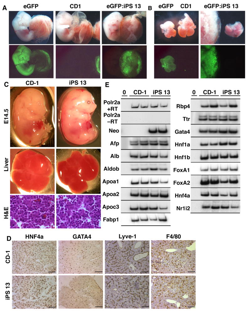
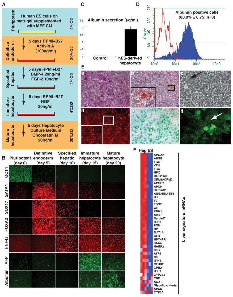
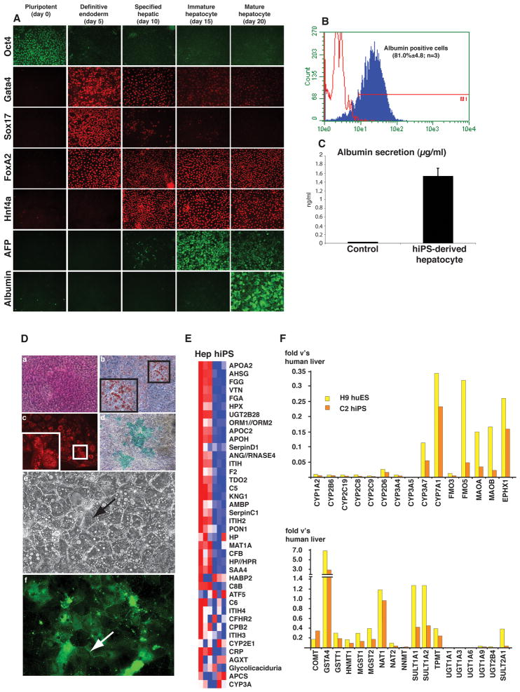
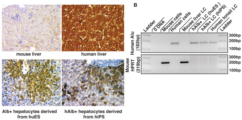
Conclusion: We demonstrate that mouse iPS cells retain full potential for fetal liver development and describe a procedure that facilitates the efficient generation of highly differentiated human hepatocyte-like cells from iPS cells that display key liver functions and can integrate into the hepatic parenchyma in vivo.


Figures

Figure 1
Figure 1. Fetal livers derived from mouse iPS cells
A) Brightfield (top) and fluorescent (bottom) images of CAG-eGFP+/− (eGFP), eGFP negative (CD1), and iPS cell–derived (eGFP:iPS 13) E12.5 embryos. B) Brightfield and fluorescent images of CAG-eGFP+/−(eGFP;left), eGFP-negative (CD1;right), and iPS cell-derived E12.5 fetal livers (eGFP:iPS 13). C) Whole mount images of E14.5 embryos (top) and their livers (middle) derived from wild-type (CD-1) and iPS cells (iPS 13). Bottom panels show hematoxylin and eosin stained sections through control (CD-1) and iPS cell–derived (iPS 13) E14.5 livers. D) Immunohistochemistry revealing marker expression in hepatocytes (HNF4a), endothelial cells (GATA4), sinusoidal cells (Lyve-1), and macrophages/Kupffer cells (F4/80) in control (CD-1) and iPS cell–derived (iPS 13) E14.5 livers. Scale bars = 100μm. E) RT-PCR analyses of two control (CD-1) and iPS–derived (iPS 13) E14.5 livers showing steady levels of characteristic liver mRNAs as well as neomycin phosphotransferase II mRNA, which is only expressed in iPS cell-derived livers. RNA polymerase II mRNA (Pol2ra) was used as a loading control, and reactions without reverse transcriptase (−RT) or template (0) confirmed the absence of contaminating DNA.
Figure 2
Figure 2. Generation of hepatocytes from human ES cells
A) Flow diagram outlining the procedure used to control hepatocyte differentiation. B) Hepatocyte differentiation from huES cells was monitored by immunocytochemistry at day 0, 5, 10, 15, and 20 using antibodies that recognized OCT3/4, FOXA2, SOX17, GATA4, HNF4a, Alphafetoprotein (AFP), and Albumin. Results are representative of three independent differentiation experiments. C) Albumin secretion by huES cell–derived hepatocytes was identified after 3–days of culture in medium by ELISA. D) Representative flow cytometry profile showing the average number of Albumin positive hepatocytes in three independent differentiation experiments = 80.9%±0.75. E) hES cell–derived hepatocytes were shown to store glycogen by PAS staining (a); store lipids by Oil Red O staining (b); to display hepatocyte morphology including bi-nucleated cells (black arrow) (c); efficiently uptake LDL using Dil-LDL (d); metabolize indocyanine green (e); and localize dichlorofluorescein diacetate to their plasma membranes (white arrow) (f). F) Heat map of gene array analyses demonstrating that a series of 40 mRNAs that are solely expressed in livers were increased (red=high, blue=low) following differentiation (Hep) compared with undifferentiated (ES) cells.
Figure 3
Figure 3. Differentiation of hepatocytes from human iPS cells
A) Hepatocyte differentiation from hiPS cells was followed by detecting OCT3/4, FOXA2, SOX17, GATA4, HNF4a, Alphafetoprotein (AFP), and Albumin (ALB) by immunocytochemistry at day 0, 5, 10, 15, and 20 days. Results are representative of three independent differentiation experiments. B) Representative flow cytometry profile showing the average number of Albumin positive hiPS–derived hepatocytes in three independent differentiation experiments = 81.0%±4.8. C) Albumin secretion by hiPS cell–derived hepatocytes was identified in the medium after 3–days of culture using ELISA. D) Hepatocyte–like cells derived from hiPS cells were shown to store glycogen by PAS staining (a); store lipids by Oil Red O staining (b); uptake LDL using Dil-LDL (c); and metabolize indocyanine green (d); have similar morphology to hepatocytes with some cells being bi-nucleated (black arrow) (e); and direct dichlorofluorescein diacetate to plasma membranes (white arrow) (f). E) Heat map of gene array analyses showing that expression of 40 liver–specific mRNAs was increased (red) following differentiation (Hep) compared with undifferentiated (hiPS) cells where the majority of these mRNAs were expressed at relatively low levels (blue). F) Bar graphs showing the levels of mRNAs, determined by real-time qRT-PCR, encoding Phase I and II enzymes in hepatocyte–like cells derived from H9 huES cells (yellow) and C2 hiPS cells (orange) expressed as fold of levels found in cadaveric human liver samples.
Figure 4
Figure 4. Integration of huES and hiPS cell–derived hepatocytes into the mouse hepatic parenchyma
A) Micrographs showing the identification of cells expressing human Albumin (brown) in human livers (upper right) and in mouse livers injected with huES cell– (lower left) and hiPS cell–derived (lower right) hepatocytes but not in uninjected control mouse livers (top right). B) PCR analyses using primers that specifically recognize human Alu or mouse HPRT sequences on genomic DNA isolated from control mouse and human fibroblasts as well as cells collected by laser capture from sections through mouse liver, human tonsil, and albumin positive cells from mouse livers injected with huES cell– or hiPS cell–derived hepatocyes. Amplifications performed without template ensured the absence of contaminating DNA.

Comment in

Similar articles

Cited by

References

    1. Lowry WE, Richter L, Yachechko R, et al. Generation of human induced pluripotent stem cells from dermal fibroblasts. Proc Natl Acad Sci U S A. 2008;105:2883–2888. - PMC - PubMed
    1. Park IH, Zhao R, West JA, et al. Reprogramming of human somatic cells to pluripotency with defined factors. Nature. 2008;451:141–146. - PubMed
    1. Takahashi K, Tanabe K, Ohnuki M, et al. Induction of pluripotent stem cells from adult human fibroblasts by defined factors. Cell. 2007;131:861–872. - PubMed
    1. Takahashi K, Yamanaka S. Induction of pluripotent stem cells from mouse embryonic and adult fibroblast cultures by defined factors. Cell. 2006;126:663–676. - PubMed
    1. Yu J, Vodyanik MA, Smuga-Otto K, et al. Induced pluripotent stem cell lines derived from human somatic cells. Science. 2007;318:1917–1920. - PubMed

Publication types