

Confocal laser endomicroscopy in the "in vivo" histological diagnosis of the gastrointestinal tract
- PMID: 19998496
- PMCID: PMC2791268
- DOI: 10.3748/wjg.15.5770
Confocal laser endomicroscopy in the "in vivo" histological diagnosis of the gastrointestinal tract
Abstract
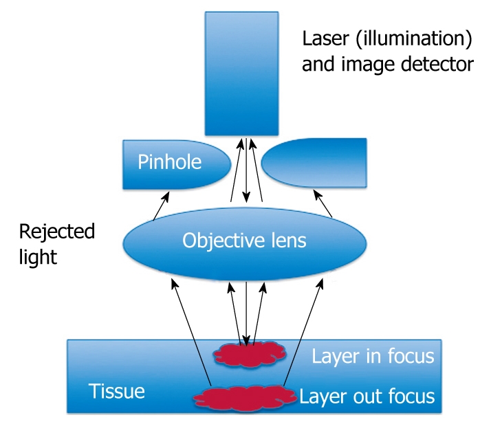
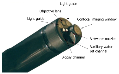
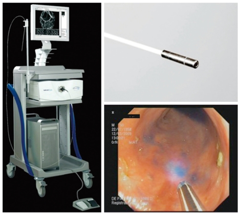
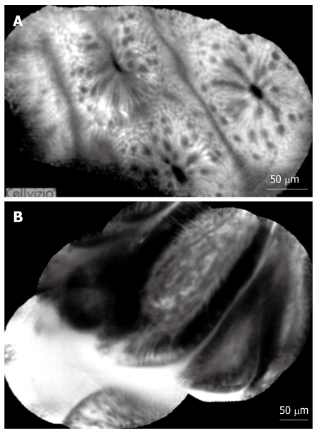
Recent technological advances in miniaturization have allowed for a confocal scanning microscope to be integrated into a conventional flexible endoscope, or into trans-endoscopic probes, a technique now known as confocal endomicroscopy or confocal laser endomicroscopy. This newly-developed technology has enabled endoscopists to collect real-time in vivo histological images or "virtual biopsies" of the gastrointestinal mucosa during endoscopy, and has stimulated significant interest in the application of this technique in clinical gastroenterology. This review aims to evaluate the current data on the technical aspects and the utility of this new technology in clinical gastroenterology and its potential impact in the future, particularly in the screening or surveillance of gastrointestinal neoplasia.
Figures
Similar articles
-
Molecular confocal laser endomicroscopy: a novel technique for in vivo cellular characterization of gastrointestinal lesions.World J Gastroenterol. 2014 Jun 28;20(24):7794-800. doi: 10.3748/wjg.v20.i24.7794. World J Gastroenterol. 2014. PMID: 24976717 Free PMC article.
-
Feasibility of confocal endomicroscopy in the diagnosis of pediatric gastrointestinal disorders.World J Gastroenterol. 2009 May 14;15(18):2214-9. doi: 10.3748/wjg.15.2214. World J Gastroenterol. 2009. PMID: 19437560 Free PMC article.
-
Current application of confocal endomicroscopy in gastrointestinal disorders.J Gastroenterol Hepatol. 2008 Oct;23(10):1483-91. doi: 10.1111/j.1440-1746.2008.05469.x. Epub 2008 Aug 28. J Gastroenterol Hepatol. 2008. PMID: 18761561 Review.
-
Status of confocal laser endomicroscopy in gastrointestinal disease.Trop Gastroenterol. 2012 Jan-Mar;33(1):9-20. doi: 10.7869/tg.2012.3. Trop Gastroenterol. 2012. PMID: 22803291 Review.
-
Comparison between two types of confocal laser endomicroscopy in gastrointestinal tract.J Dig Dis. 2015 May;16(5):279-85. doi: 10.1111/1751-2980.12245. J Dig Dis. 2015. PMID: 25762057 Clinical Trial.
Cited by
-
Chronic radiation proctitis: tricks to prevent and treat.Int J Colorectal Dis. 2015 Oct;30(10):1293-303. doi: 10.1007/s00384-015-2289-4. Epub 2015 Jul 23. Int J Colorectal Dis. 2015. PMID: 26198994 Free PMC article. Review.
-
Endomicroscopy with Fluorescent CD105 Antibodies for "In Vivo" Imaging of Colorectal Cancer Angiogenesis.Curr Health Sci J. 2015 Jul-Sep;41(3):288-292. doi: 10.12865/CHSJ.41.03.17. Epub 2015 Mar 15. Curr Health Sci J. 2015. PMID: 30538832 Free PMC article.
-
Capsule optoacoustic endoscopy for esophageal imaging.J Biophotonics. 2019 Oct;12(10):e201800439. doi: 10.1002/jbio.201800439. Epub 2019 Jun 25. J Biophotonics. 2019. PMID: 31034135 Free PMC article.
-
Confocal laser endomicroscopy and immunoendoscopy for real-time assessment of vascularization in gastrointestinal malignancies.World J Gastroenterol. 2011 Jan 7;17(1):21-7. doi: 10.3748/wjg.v17.i1.21. World J Gastroenterol. 2011. PMID: 21218080 Free PMC article. Review.
-
Probe-based confocal laser endomicroscopy for in vivo evaluation of the tumor vasculature in gastric and rectal carcinomas.Sci Rep. 2017 Aug 29;7(1):9819. doi: 10.1038/s41598-017-10963-1. Sci Rep. 2017. PMID: 28852161 Free PMC article.
References
-
- Polglase AL, McLaren WJ, Skinner SA, Kiesslich R, Neurath MF, Delaney PM. A fluorescence confocal endomicroscope for in vivo microscopy of the upper- and the lower-GI tract. Gastrointest Endosc. 2005;62:686–695. - PubMed
-
- Kiesslich R, Goetz M, Neurath MF. Confocal laser endomicroscopy for gastrointestinal diseases. Gastrointest Endosc Clin N Am. 2008;18:451–466, viii. - PubMed
-
- Kantsevoy SV, Adler DG, Conway JD, Diehl DL, Farraye FA, Kaul V, Kethu SR, Kwon RS, Mamula P, Rodriguez SA, et al. Confocal laser endomicroscopy. Gastrointest Endosc. 2009;70:197–200. - PubMed
Publication types
MeSH terms
Substances
LinkOut - more resources
Full Text Sources
Other Literature Sources
Medical
