

Role of diffusion-weighted magnetic resonance imaging in the differential diagnosis of focal hepatic lesions
- PMID: 19998501
- PMCID: PMC2791273
- DOI: 10.3748/wjg.15.5805
Role of diffusion-weighted magnetic resonance imaging in the differential diagnosis of focal hepatic lesions
Abstract
Aim: To evaluate the utility of diffusion-weighted imaging (DWI) in screening and differential diagnosis of benign and malignant focal hepatic lesions.
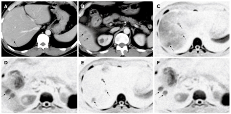
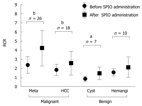
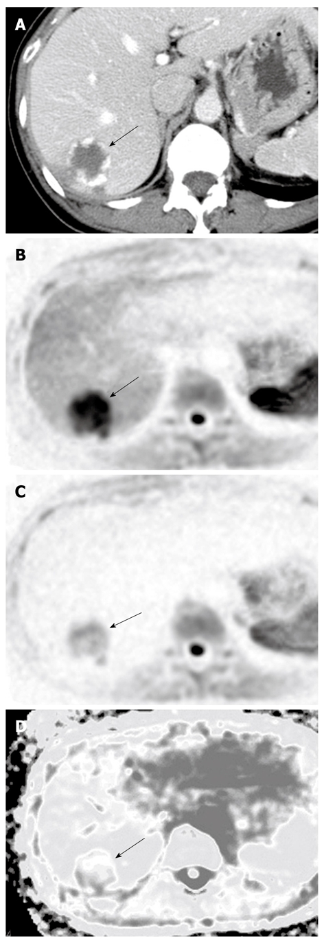
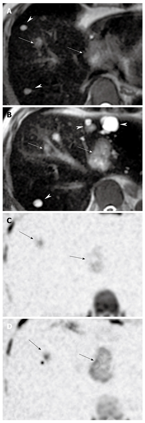
Methods: Magnetic resonance imaging (MRI) examinations were performed using the Signa Excite Xl Twin Speed 1.5T system (GE Healthcare, Milwaukee, WI, USA). Seventy patients who had undergone MRI of the liver [29 hepatocellular carcinomas (HCC), four cholangiocarcinomas, 34 metastatic liver cancers, 10 hemangiomas, and eight cysts] between April 2004 and August 2008 were retrospectively evaluated. Visualization of lesions, relative contrast ratio (RCR), and apparent diffusion coefficient (ADC) were compared between benign and malignant lesions on DWI. Superparamagnetic iron oxide (SPIO) was administered to 59 patients, and RCR was compared pre- and post-administration.
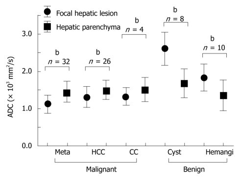
Results: DWI showed higher contrast between malignant lesions (especially in multiple small metastatic cancers) and surrounding liver parenchyma than did contrast-enhanced computed tomography. ADCs (mean +/- SD x 10(-3) mm(2)/s) were significantly lower (P < 0.05) in malignant lesions (HCC: 1.31 +/- 0.28 and liver metastasis: 1.11 +/- 0.22) and were significantly higher in benign lesions (hemangioma: 1.84 +/- 0.37 and cyst: 2.61 +/- 0.45) than in the surrounding hepatic tissues. RCR between malignant lesions and surrounding hepatic tissues significantly improved after SPIO administration, but RCRs in benign lesions were not improved.
Conclusion: DWI is a simple and sensitive method for screening focal hepatic lesions and is useful for differential diagnosis.
Figures
Similar articles
-
Diagnostic value of diffusion weighted MRI and ADC in differential diagnosis of cavernous hemangioma of the liver.Afr Health Sci. 2016 Mar;16(1):227-33. doi: 10.4314/ahs.v16i1.30. Afr Health Sci. 2016. PMID: 27358636 Free PMC article.
-
Effect of intravenous gadolinium-DTPA on diffusion-weighted magnetic resonance images for evaluation of focal hepatic lesions.J Comput Assist Tomogr. 2005 Mar-Apr;29(2):176-80. doi: 10.1097/01.rct.0000157472.98277.5c. J Comput Assist Tomogr. 2005. PMID: 15772533
-
[DWI MAGNETIC RESONANCE IN CHARACTERIZATION OF FOCAL LIVER LESIONS].Acta Med Croatica. 2016 Sep;70(3):179-84. Acta Med Croatica. 2016. PMID: 29064209 Croatian.
-
Lesion discrimination with breath-hold hepatic diffusion-weighted imaging: a meta-analysis.World J Gastroenterol. 2015 Feb 7;21(5):1621-7. doi: 10.3748/wjg.v21.i5.1621. World J Gastroenterol. 2015. PMID: 25663782 Free PMC article. Review.
-
Diffusion weighted imaging in the liver.World J Gastroenterol. 2010 Apr 7;16(13):1567-76. doi: 10.3748/wjg.v16.i13.1567. World J Gastroenterol. 2010. PMID: 20355235 Free PMC article. Review.
Cited by
-
Role of the apparent diffusion coefficient measurement by diffusion weighted magnetic resonance imaging in the diagnosis of Fasciola hepatica in the liver.Jpn J Radiol. 2011 Dec;29(10):688-94. doi: 10.1007/s11604-011-0614-6. Epub 2011 Oct 19. Jpn J Radiol. 2011. PMID: 22009419
-
Diffusion-weighted whole-body imaging with background suppression (DWIBS) is effective and economical for detection of metastasis or recurrence of lung cancer.Thorac Cancer. 2021 Mar;12(5):676-684. doi: 10.1111/1759-7714.13820. Epub 2021 Jan 21. Thorac Cancer. 2021. PMID: 33476488 Free PMC article.
-
Whole-Lesion Apparent Diffusion Coefficient Histogram Analysis: Significance for Discriminating Lung Cancer from Pulmonary Abscess and Mycobacterial Infection.Cancers (Basel). 2021 May 31;13(11):2720. doi: 10.3390/cancers13112720. Cancers (Basel). 2021. PMID: 34072867 Free PMC article.
-
Effect of imaging parameters on the accuracy of apparent diffusion coefficient and optimization strategies.Diagn Interv Radiol. 2016 Jan-Feb;22(1):101-7. doi: 10.5152/dir.2015.14440. Diagn Interv Radiol. 2016. PMID: 26573977 Free PMC article.
-
Comparison of DWIBS/T2 image fusion and PET/CT for the diagnosis of cancer in the abdominal cavity.Exp Ther Med. 2017 Oct;14(4):3754-3760. doi: 10.3892/etm.2017.4987. Epub 2017 Aug 22. Exp Ther Med. 2017. PMID: 29042975 Free PMC article.
References
-
- Le Bihan D, Breton E, Lallemand D, Aubin ML, Vignaud J, Laval-Jeantet M. Separation of diffusion and perfusion in intravoxel incoherent motion MR imaging. Radiology. 1988;168:497–505. - PubMed
-
- Sorensen AG, Buonanno FS, Gonzalez RG, Schwamm LH, Lev MH, Huang-Hellinger FR, Reese TG, Weisskoff RM, Davis TL, Suwanwela N, et al. Hyperacute stroke: evaluation with combined multisection diffusion-weighted and hemodynamically weighted echo-planar MR imaging. Radiology. 1996;199:391–401. - PubMed
-
- Warach S, Chien D, Li W, Ronthal M, Edelman RR. Fast magnetic resonance diffusion-weighted imaging of acute human stroke. Neurology. 1992;42:1717–1723. - PubMed
-
- Ichikawa T, Haradome H, Hachiya J, Nitatori T, Araki T. Diffusion-weighted MR imaging with single-shot echo-planar imaging in the upper abdomen: preliminary clinical experience in 61 patients. Abdom Imaging. 1999;24:456–461. - PubMed
MeSH terms
LinkOut - more resources
Full Text Sources
Medical
Miscellaneous
