Iodized oil uptake assessment with cone-beam CT in chemoembolization of small hepatocellular carcinomas
- PMID: 19998505
- PMCID: PMC2791277
- DOI: 10.3748/wjg.15.5833
Iodized oil uptake assessment with cone-beam CT in chemoembolization of small hepatocellular carcinomas
Abstract
Aim: To evaluate the utility of assessing iodized oil uptake with cone-beam computed tomography (CT) in transarterial chemoembolization (TACE) for small hepatocellular carcinoma (HCC).
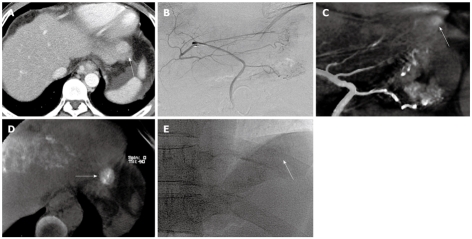
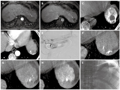
Methods: Cone-beam CT provided by a biplane flat-panel detector angiography suite was performed on eighteen patients (sixteen men and two women; 41-76 years; mean age, 58.9 years) directly after TACE for small HCC (26 nodules under 30 mm; mean diameter, 11.9 mm; range, 5-28 mm). The pre-procedural locations of the tumors were evaluated using triphasic multi-detector row helical computed tomography (MDCT). The tumor locations on MDCT and the iodized oil uptake by the tumors were analyzed on cone-beam CT and on spot image directly after the procedures.
Results: All lesions on preprocedural MDCT were detected using iodized oil uptake in the lesions on cone-beam CT (sensitivity 100%, 26/26). Spot image depicted iodized oil uptake in 22 of the lesions (sensitivity 85%). The degree of iodized oil uptake was overestimated (9%, 2/22) or underestimated (14%, 3/22) on spot image in five nodules compared with that of cone-beam CT.
Conclusion: Cone-beam CT is a useful and convenient tool for assessing the iodized oil uptake of small hepatic tumors (< 3 cm) directly after TACE.
Figures


Similar articles
-
The usefulness of cone-beam computed tomography during chemoembolization of hepatocellular carcinomas fed exclusively by the cystic artery.Jpn J Radiol. 2016 Nov;34(11):747-753. doi: 10.1007/s11604-016-0580-0. Epub 2016 Sep 21. Jpn J Radiol. 2016. PMID: 27653751
-
Usefulness of cone-beam volume CT with flat panel detectors in conjunction with catheter angiography for transcatheter arterial embolization.J Vasc Interv Radiol. 2007 Dec;18(12):1508-16. doi: 10.1016/j.jvir.2007.08.003. J Vasc Interv Radiol. 2007. PMID: 18057285
-
C-arm CT for assessing initial failure of iodized oil accumulation in chemoembolization of hepatocellular carcinoma.AJR Am J Roentgenol. 2011 Aug;197(2):W337-42. doi: 10.2214/AJR.10.5614. AJR Am J Roentgenol. 2011. PMID: 21785062
-
Efficacy of cone-beam computed tomography during transcatheter arterial chemoembolization for hepatocellular carcinoma.Jpn J Radiol. 2011 Jul;29(6):371-7. doi: 10.1007/s11604-011-0568-8. Epub 2011 Jul 24. Jpn J Radiol. 2011. PMID: 21786092 Review.
-
Initiative on Superselective Conventional Transarterial Chemoembolization Results (INSPIRE).Cardiovasc Intervent Radiol. 2022 Oct;45(10):1430-1440. doi: 10.1007/s00270-022-03233-9. Epub 2022 Aug 17. Cardiovasc Intervent Radiol. 2022. PMID: 35978174 Free PMC article. Review.
Cited by
-
The Role of Immediate Post-Procedural Cone-Beam Computed Tomography (CBCT) in Predicting the Early Radiologic Response of Hepatocellular Carcinoma (HCC) Nodules to Drug-Eluting Bead Transarterial Chemoembolization (DEB-TACE).J Clin Med. 2022 Nov 29;11(23):7089. doi: 10.3390/jcm11237089. J Clin Med. 2022. PMID: 36498664 Free PMC article.
-
Quantitative assessment of lipiodol deposition after chemoembolization: comparison between cone-beam CT and multidetector CT.J Vasc Interv Radiol. 2013 Dec;24(12):1837-44. doi: 10.1016/j.jvir.2013.08.017. Epub 2013 Oct 1. J Vasc Interv Radiol. 2013. PMID: 24094672 Free PMC article.
-
Lipiodol retention pattern assessed by cone beam computed tomography during conventional transarterial chemoembolization of hepatocellular carcinoma: accuracy and correlation with response.Cancer Imaging. 2016 Oct 3;16(1):32. doi: 10.1186/s40644-016-0090-4. Cancer Imaging. 2016. PMID: 27716376 Free PMC article.
-
Novel imaging biomarkers of response to transcatheter arterial chemoembolization in hepatocellular carcinoma patients.Chin J Cancer Res. 2015 Dec;27(6):611-26. doi: 10.3978/j.issn.1000-9604.2015.07.02. Chin J Cancer Res. 2015. PMID: 26752936 Free PMC article. No abstract available.
-
Radiation dose to procedural personnel and patients from an X-ray volume imaging system.Eur Radiol. 2013 Dec;23(12):3262-70. doi: 10.1007/s00330-013-2939-2. Epub 2013 Jun 29. Eur Radiol. 2013. PMID: 23812243 Clinical Trial.
References
-
- Murakami T, Kim T, Takamura M, Hori M, Takahashi S, Federle MP, Tsuda K, Osuga K, Kawata S, Nakamura H, et al. Hypervascular hepatocellular carcinoma: detection with double arterial phase multi-detector row helical CT. Radiology. 2001;218:763–767. - PubMed
-
- Kim SK, Lim JH, Lee WJ, Kim SH, Choi D, Lee SJ, Lim HK, Kim H. Detection of hepatocellular carcinoma: comparison of dynamic three-phase computed tomography images and four-phase computed tomography images using multidetector row helical computed tomography. J Comput Assist Tomogr. 2002;26:691–698. - PubMed
-
- Bartolozzi C, Lencioni R, Caramella D, Palla A, Bassi AM, Di Candio G. Small hepatocellular carcinoma. Detection with US, CT, MR imaging, DSA, and Lipiodol-CT. Acta Radiol. 1996;37:69–74. - PubMed
-
- De Santis M, Romagnoli R, Cristani A, Cioni G, Casolo A, Vici FF, Ventura E. MRI of small hepatocellular carcinoma: comparison with US, CT, DSA, and Lipiodol-CT. J Comput Assist Tomogr. 1992;16:189–197. - PubMed
Publication types
MeSH terms
Substances
LinkOut - more resources
Full Text Sources
Medical
Miscellaneous