

Changes in intestinal mucosal immune barrier in rats with endotoxemia
- PMID: 19998507
- PMCID: PMC2791279
- DOI: 10.3748/wjg.15.5843
Changes in intestinal mucosal immune barrier in rats with endotoxemia
Abstract
Aim: To investigate the dysfunction of the immunological barrier of the intestinal mucosa during endotoxemia and to elucidate the potential mechanism of this dysfunction.
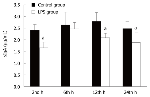
Methods: Male Wistar rats were randomly distributed into two groups: control group and lipopolysaccharide (LPS) group. Endotoxemia was induced by a single caudal venous injection of LPS. Animals were sacrificed in batches 2, 6, 12 and 24 h after LPS infusion. The number of microfold (M)-cells, dendritic cells (DCs), CD4(+) T cells, CD8(+) T cells, regulatory T (Tr) cells and IgA(+) B cells in the intestinal mucosa were counted after immunohistochemical staining. Apoptotic lymphocytes were counted after TUNEL staining. The levels of interleukin (IL)-4, interferon (IFN)-gamma and forkhead box P3 (Foxp3) in mucosal homogenates were measured by ELISA. The secretory IgA (sIgA) content in the total protein of one milligram of small intestinal mucus was detected using a radioimmunological assay.
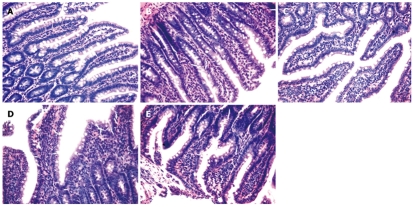
Results: This research demonstrated that LPS-induced endotoxemia results in small intestinal mucosa injury. The number of M-cells, DCs, CD8(+) T cells, and IgA(+) B cells were decreased while Tr cell and apoptotic lymphocyte numbers were increased significantly. The number of CD4(+) T cells increased in the early stages and then slightly decreased by 24 h. The level of IL-4 significantly increased in the early stages and then reversed by the end of the study period. The level of IFN-gamma increased slightly in the early stages and then decreased markedly by the 24 h time point. Level of Foxp3 increased whereas sIgA level decreased.
Conclusion: Mucosal immune dysfunction forms part of the intestinal barrier injury during endotoxemia. The increased number and function of Tr cells as well as lymphocyte apoptosis result in mucosal immunodeficiency.
Figures


Similar articles
-
[Effect of Madecassoside on Intestinal Mucosal Immunity in Collagen-Induced Arthritis Rats].Zhong Yao Cai. 2015 Feb;38(2):333-8. Zhong Yao Cai. 2015. PMID: 26415412 Chinese.
-
[Effects of astragalus polysaccharide on intestinal immune function of rats with severe scald injury].Zhonghua Shao Shang Za Zhi. 2015 Feb;31(1):30-6. Zhonghua Shao Shang Za Zhi. 2015. PMID: 25876637 Chinese.
-
Decreased IgA+ plasma cells and IgA expression in acute liver necrosis mice.World J Gastroenterol. 2010 Aug 14;16(30):3827-33. doi: 10.3748/wjg.v16.i30.3827. World J Gastroenterol. 2010. PMID: 20698046 Free PMC article.
-
Gut Microbiome Homeostasis and the CD4 T- Follicular Helper Cell IgA Axis in Human Immunodeficiency Virus Infection.Front Immunol. 2021 Mar 19;12:657679. doi: 10.3389/fimmu.2021.657679. eCollection 2021. Front Immunol. 2021. PMID: 33815419 Free PMC article. Review.
-
Re-thinking the functions of IgA(+) plasma cells.Gut Microbes. 2014;5(5):652-62. doi: 10.4161/19490976.2014.969977. Gut Microbes. 2014. PMID: 25483334 Free PMC article. Review.
Cited by
-
Visfatin Regulates Inflammatory Mediators in Mouse Intestinal Mucosa Through Toll-Like Receptors Signaling Under Lipopolysaccharide Stress.Arch Immunol Ther Exp (Warsz). 2021 Apr 15;69(1):11. doi: 10.1007/s00005-021-00611-y. Arch Immunol Ther Exp (Warsz). 2021. PMID: 33856572
-
Shenfu injection prolongs survival and protects the intestinal mucosa in rats with sepsis by modulating immune response.Turk J Gastroenterol. 2019 Apr;30(4):364-371. doi: 10.5152/tjg.2019.18418. Turk J Gastroenterol. 2019. PMID: 30666971 Free PMC article.
-
Lipopolysaccharide-Induced Increase in Intestinal Permeability Is Mediated by TAK-1 Activation of IKK and MLCK/MYLK Gene.Am J Pathol. 2019 Apr;189(4):797-812. doi: 10.1016/j.ajpath.2018.12.016. Epub 2019 Feb 1. Am J Pathol. 2019. PMID: 30711488 Free PMC article.
-
Markers of inflammation and coagulation may be modulated by enteral feeding strategy.JPEN J Parenter Enteral Nutr. 2012 Nov;36(6):732-40. doi: 10.1177/0148607111433054. Epub 2012 Feb 7. JPEN J Parenter Enteral Nutr. 2012. PMID: 22318965 Free PMC article. Clinical Trial.
-
Chronic bile duct hyperplasia is a chronic graft dysfunction following liver transplantation.World J Gastroenterol. 2012 Mar 14;18(10):1038-47. doi: 10.3748/wjg.v18.i10.1038. World J Gastroenterol. 2012. PMID: 22416178 Free PMC article.
References
-
- Angus DC, Linde-Zwirble WT, Lidicker J, Clermont G, Carcillo J, Pinsky MR. Epidemiology of severe sepsis in the United States: analysis of incidence, outcome, and associated costs of care. Crit Care Med. 2001;29:1303–1310. - PubMed
-
- Cheng B, Xie G, Yao S, Wu X, Guo Q, Gu M, Fang Q, Xu Q, Wang D, Jin Y, et al. Epidemiology of severe sepsis in critically ill surgical patients in ten university hospitals in China. Crit Care Med. 2007;35:2538–2546. - PubMed
-
- Brun-Buisson C. [Epidemiology of severe sepsis] Presse Med. 2006;35:513–520. - PubMed
-
- Opal SM, Scannon PJ, Vincent JL, White M, Carroll SF, Palardy JE, Parejo NA, Pribble JP, Lemke JH. Relationship between plasma levels of lipopolysaccharide (LPS) and LPS-binding protein in patients with severe sepsis and septic shock. J Infect Dis. 1999;180:1584–1589. - PubMed
Publication types
MeSH terms
Substances
LinkOut - more resources
Full Text Sources
Research Materials
Miscellaneous
