. 2010 Jan 11;11(1):192-200.
doi: 10.1021/bm9010672.

Quantitative Correlation between the protein primary sequences and secondary structures in spider dragline silks

Affiliations

Quantitative Correlation between the protein primary sequences and secondary structures in spider dragline silks

Janelle E Jenkins et al. Biomacromolecules. .

Abstract

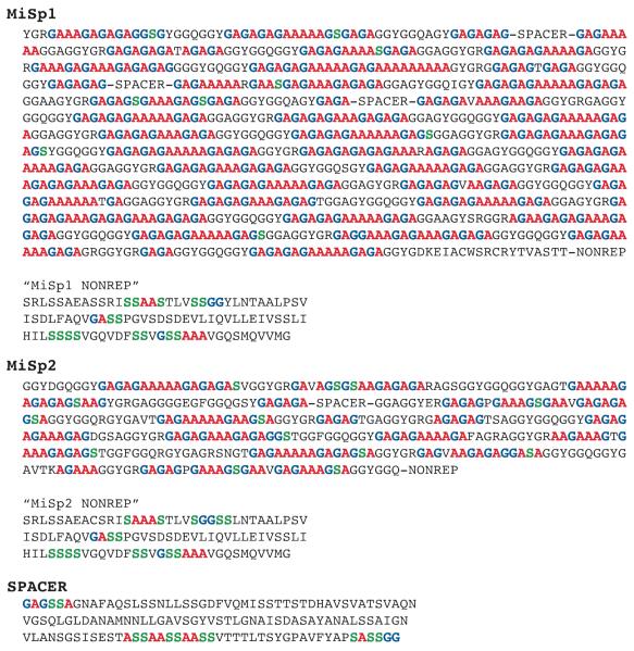
Synthetic spider silk holds great potential for use in various applications spanning medical uses to ultra lightweight armor; however, producing synthetic fibers with mechanical properties comparable to natural spider silk has eluded the scientific community. Natural dragline spider silks are commonly made from proteins that contain highly repetitive amino acid motifs, adopting an array of secondary structures. Before further advances can be made in the production of synthetic fibers based on spider silk proteins, it is imperative to know the percentage of each amino acid in the protein that forms a specific secondary structure. Linking these percentages to the primary amino acid sequence of the protein will establish a structural foundation for synthetic silk. In this study, nuclear magnetic resonance (NMR) techniques are used to quantify the percentage of Ala, Gly, and Ser that form both beta-sheet and helical secondary structures. The fraction of these three amino acids and their secondary structure are quantitatively correlated to the primary amino acid sequence for the proteins that comprise major and minor ampullate silk from the Nephila clavipes spider providing a blueprint for synthetic spider silks.


Figures

Figure 1
Figure 1
13C-13C correlation NMR experiments of N. clavipes Mi silk with DARR mixing times of (A) 150 ms and (B) 1 s. Projections are taken from the 1 s DARR experiment at the (C, top) 31-helical Ala Cβ (17.4 ppm), (C, middle) β-sheet Ala Cβ (21.7 ppm), and (C, bottom) Gly Cα (43.4 ppm). * Indicates weak intermolecular correlations between Gly and Ala.
Figure 2
Figure 2
13C-13C through-bond double quantum/single quantum (DQ/SQ) correlation collected with refocused INADEQUATE NMR experiments of N. clavipes Mi silk: (A) An initial cross polarization step was used to enhance carbon magnetization for the dry silk, while a direct carbon INADEQUATE was used for the water wetted silk (B & C). A recycle delay of (B) 1 s enhances mobile components that contain shorter T1 relaxation times, while a (C) 3 s recycle delay emphasizes rigid components. Carbonyl regions are blown up in insets to reveal two distinct CO resonances for Ala and Gly.
Figure 3
Figure 3
Carbonyl region from fully relaxed 13C DD-MAS NMR spectrum of water wetted N. clavipes (A) Ma silk and (B) Mi silk. The spectra were fit to extract the percent of Gly and Ala that each adopt both β-sheet and helical conformations (see Table 2). The original data is in black, the sum of the fits are in grey, and the individual fits are dotted lines.
Figure 4
Figure 4
Aliphatic region from fully relaxed 13C DD-MAS NMR spectrum of water wetted N. clavipes (A) Ma and (B) Mi silk. The Ser Cβ fit was used to determine the percentage of Ser incorporated into a β-sheet or helical conformation. The Ala Cβ in both the Ma and Mi silk were fit and compared to carbonyl fits. This provided a self-consistent check for the percentage of Ala in a β-sheet or helical conformation extracted from the carbonyl fits. The original data is in black, the sum of the fits are in grey, and the individual fits are dotted lines.
Figure 5
Figure 5
Primary amino acid sequences for N. clavipes MaSp1 and MaSp2, the two proteins that make up Ma silk. Colored Ala (red), Gly (blue), and Ser (green) represent β-sheet structure. The fraction of Ala, Gly, and Ser in a β-sheet structure in silk predicted by the primary amino acid sequences are 86%, 26%, and 19%, respectively. Percentage of β-sheet determined experimentally by NMR agrees with the above structural model.
Figure 6
Figure 6
Primary amino acid sequences for N. clavipes MiSp1, MiSp2, the Ser-rich nonrepetitive spacer region (“spacer”), and carboxy-terminal nonrepetitive (“nonrep”) regions that make up Mi silk. Colored Ala (red), Gly (blue), and Ser (green) represent β-sheet structure. The predicted percentage of Ala, Gly, and Ser that form β-sheet in Mi silk (75-83%, 44-52%, and 41-48%, respectively) agrees with experimental NMR data. The ratio of MiSp1 to MiSp2 is unknown, therefore a range in percentage predicted by the AA sequence is based on 100% MiSp1 or MiSp2.

Similar articles

Cited by

References

    1. Shear WA, Palmer JM, Coddington JA, Bonamo PM. Science. 1989;246:479–481. - PubMed
    1. Lewis R. Chem. Rev. 2006;106:3762–3774. - PubMed
    1. Vollrath F, Porter D. Soft Matter. 2006;2:377–385. - PubMed
    1. Gosline J, DeMont E, Denny M. Endeavour. 1986;10:37–43.
    1. Swanson BO, Blackledge TA, Beltran J, Hayashi CY. Appl. Phys. A. 2006;82:213–218.

Publication types