. 2010 May;16(5):1469-77.
doi: 10.1089/ten.TEA.2009.0118.

Principles of biomimetic vascular network design applied to a tissue-engineered liver scaffold

Affiliations

Principles of biomimetic vascular network design applied to a tissue-engineered liver scaffold

David M Hoganson et al. Tissue Eng Part A. 2010 May.

Abstract

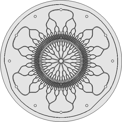
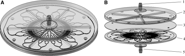
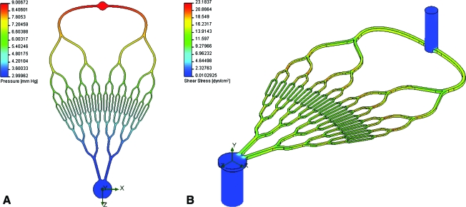
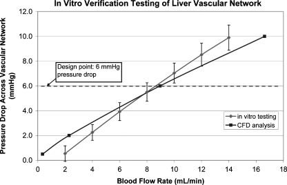
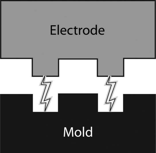
Branched vascular networks are a central component of scaffold architecture for solid organ tissue engineering. In this work, seven biomimetic principles were established as the major guiding technical design considerations of a branched vascular network for a tissue-engineered scaffold. These biomimetic design principles were applied to a branched radial architecture to develop a liver-specific vascular network. Iterative design changes and computational fluid dynamic analysis were used to optimize the network before mold manufacturing. The vascular network mold was created using a new mold technique that achieves a 1:1 aspect ratio for all channels. In vitro blood flow testing confirmed the physiologic hemodynamics of the network as predicted by computational fluid dynamic analysis. These results indicate that this biomimetic liver vascular network design will provide a foundation for developing complex vascular networks for solid organ tissue engineering that achieve physiologic blood flow.


Figures

FIG. 1.
FIG. 1.
Top view of radial branching vascular network designed according to biomimetic principles. The six radial vascular inlets are shown at the periphery, and the vascular channels bifurcate into several generations of inlet channels toward the smallest channels. The channels then coalesce into 12 outlet channels and enter the central venous outlet.
FIG. 2.
FIG. 2.
Isometric view of single-layer liver vascular network with header and nozzles (A). Exploded view of single-layer vascular network showing inlet nozzle (1), header layer (2), spacer (3), vascular layer (4), and outlet nozzle (5) (B). Flow enters the nozzle and is divided into six channels in the header layer to enter the radial inlets of the vascular layer. Blood flows through the vascular layer and collects at the central outlet and flows out of the lower nozzle.
FIG. 3.
FIG. 3.
Pressure drop from radial inlet to central outlet of vascular network. The pressure drop is uniform across the network; due to venous scaling, more pressure drop occurs in the inlet generations than the outlet generations (A). Isometric view of the shear stress of the vascular network. Shear stress variation was minimized by utilizing both Murray's law for channel diameter relationships and channels with 1:1 aspect ratio (B). Color images available online at www.liebertonline.com/ten.
FIG. 4.
FIG. 4.
Graph of in vitro verification testing results (error bars ± one standard deviation) for blood flow compared to the CFD analysis. The error at the design point of 6 mmHg pressure drop across the network was less than 5%. CFD, computational fluid dynamic.
FIG. 5.
FIG. 5.
Electrical discharge machining utilizes an electrode of the desired pattern usually machined with a traditional milling machine in graphite. A high voltage is placed across the electrode and the steel mold material. As the electrode comes into proximity with the mold, a spark will arc across a dielectric fluid and remove a very small bit of the mold material. This process is repeated usually millions of times until the impression of the electrode has sunk into the mold to form the desired pattern.

References

    1. www.unos.org/1/8/09. http://optn.transplant.hrsa.gov/data. www.unos.org/1/8/09. http://optn.transplant.hrsa.gov/data
    1. Au P. Tam J. Fukumura D. Jain R.K. Bone marrow-derived mesenchymal stem cells facilitate engineering of long-lasting functional vasculature. Blood. 2008;111:4551. - PMC - PubMed
    1. Koike N. Fukumura D. Gralla O. Au P. Schechner J.S. Jain R.K. Tissue engineering: creation of long-lasting blood vessels. Nature. 2004;428:138. - PubMed
    1. Levenberg S. Rouwkema J. Macdonald M. Garfein E.S. Kohane D.S. Darland D.C. Marini R. van Blitterswijk C.A. Mulligan R.C. D'Amore P.A. Langer R. Engineering vascularized skeletal muscle tissue. Nat Biotechnol. 2005;23:879. - PubMed
    1. Sieminski A.L. Hebbel R.P. Gooch K.J. Improved microvascular network in vitro by human blood outgrowth endothelial cells relative to vessel-derived endothelial cells. Tissue Eng. 2005;11:1332. - PubMed

Publication types

MeSH terms

LinkOut - more resources