. 2009 Nov-Dec;17(6):380-9.
doi: 10.3109/09273940903118642.

The immune privileged retina mediates an alternative activation of J774A.1 cells

Affiliations

The immune privileged retina mediates an alternative activation of J774A.1 cells

Chun H Lau et al. Ocul Immunol Inflamm. 2009 Nov-Dec.

Abstract

Purpose: We have previously found that retinal pigment epithelial (RPE) cells suppressed endotoxin-stimulated macrophages; moreover, it induced expression of anti-inflammatory cytokines. We further assessed the possibility that the RPE is alternatively activating macrophages.

Methods: J774A.1 cells were stimulated with endotoxin and treated with the conditioned media (CM) of RPE, or neuroretinal eyecups from healthy mouse eyes. The supernatant was assayed for IL-1 beta, TNF-alpha, IL-6, IL-12(p70), and IL-10, and for nitric-oxide generation. The RPE conditioned media (RPE CM) was absorbed of known soluble factors to identify the factor that augments nitric-oxide generation.

Results: We found the RPE CM suppressed all cytokine production except IL-10, and augmented nitric-oxide generation. The augmented nitric-oxide levels were mediated by RPE derived alpha-melanocyte stimulating hormone (alpha-MSH).

Conclusions: Healthy RPE not only suppresses inflammatory activity, it promotes an alternative activation of macrophages that can further promote immune privilege.


Conflict of interest statement

Declaration of interest: The authors report no conflict of interest.

Figures

Figure 1
Figure 1
The effects of healthy retinal pigmented epithelial eye cup conditioned media (CM) on J774 macrophage cells (Mc) activated with or without endotoxin (LPS). The macrophage cells were treated with RPE CM plus 1 μg/mL of LPS and the culture supernatant was assayed after 48 h of incubation for IL-1b, TNF-a, IL-6, IL-12p70, and IL-10 using multiplex analysis. The activity of PEDF in the CM was neutralized by depleting PEDF in the CM using the absorption procedure described in the methods. The effects of five different eyecup CM were tested. Presented are mean pg/mL ± standard error of the mean for each cytokine produced by the macrophages or present in the CM. Significance is indicated by P ≤ 0.05 as indicated.
Figure 2
Figure 2
The effects of healthy neuroretina conditioned media (CM) on J774 macrophage cells (Mc) activated with or without endotoxin (LPS). The macrophage cells were treated with NR CM plus 1 μg/mL of LPS and the culture supernatant was assayed after 48 h of incubation for IL-1b, TNF-a, IL-6, IL-12p70, and IL-10 using multiplex analysis. The activity of PEDF in the CM was neutralized by depleting PEDF in the CM using the absorption procedure described in the methods. The effects of five different eyecup CM were tested. Presented are mean pg/mL ± standard error of the mean for each cytokine produced by the macrophages or present in the CM. Significance is indicated by P ≤ 0.05 as indicated.
Figure 3
Figure 3
The effects of the conditioned media (CM) on LPS-stimulated nitric oxide (NO) generation. Macrophages were stimulated with LPS and treated as described in Figure 1 and the methods with the CM from (A) RPE, (B) NR, (C) whole retinal eye cups (whole), and (D) RPE eye cups depleted of RPE cells. The 24 h macrophage culture supernatants were assayed with Griess reagent for NO. Presented are the effect of five different eyecup CM on the mean μM ± standard error of the mean of the nitrite generated by treated macrophages. Significant differences and P values are indicated between treatment groups. NS = not significant.
Figure 4
Figure 4
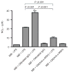
The neuropeptide a-MSH is the soluble factor in RPE CM enhanced NO production by LPS-stimulated macrophages. The experimental methods were the same as in Figure 1 for preparing the RPE CM; however, the RPE CM was depleted of a-MSH (CM[anti-a-MSH]) using the absorption procedure described in the methods. Control for the absorption procedure was done using unimmunized rabbit IgG (CM[rabbit IgG]). Presented are the results of one representative experiment repeated twice with similar relative changes in NO generation as the mean nitrite (μM) ± standard deviation from two different RPE eyecup preparations per group.

Similar articles

Cited by

References

    1. Streilein JW. Ocular immune privilege: the eye takes a dim but practical view of immunity and inflammation. J Leukoc Biol. 2003;74(2):179–185. - PubMed
    1. Taylor AW. Ocular immunosuppressive microenvironment. Chem Immunol Allergy. 2007;92:71–85. - PubMed
    1. Bok D. Evidence for an inflammatory process in age-related macular degeneration gains new support. Proc Natl Acad Sci USA. 2005;102(20):7053–7054. - PMC - PubMed
    1. Boulton M, Dayhaw-Barker P. The role of the retinal pigment epithelium: topographical variation and ageing changes. Eye. 2001;15(Pt 3):384–389. - PubMed
    1. Steinberg RH. Interactions between the retinal pigment epithelium and the neural retina. Doc Ophthalmol. 1985;60(4):327–346. - PubMed

Publication types

MeSH terms

LinkOut - more resources