. 2010 Jun;19(6):783-95.
doi: 10.1089/scd.2009.0349.

Non-cardiomyocytes influence the electrophysiological maturation of human embryonic stem cell-derived cardiomyocytes during differentiation

Affiliations

Non-cardiomyocytes influence the electrophysiological maturation of human embryonic stem cell-derived cardiomyocytes during differentiation

Changsung Kim et al. Stem Cells Dev. 2010 Jun.

Abstract

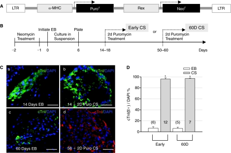
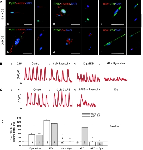
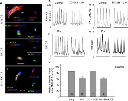
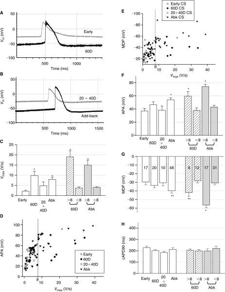
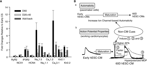
Various types of cardiomyocytes undergo changes in automaticity and electrical properties during fetal heart development. Human embryonic stem cell-derived cardiomyocytes (hESC-CMs), like fetal cardiomyocytes, are electrophysiologically immature and exhibit automaticity. We used hESC-CMs to investigate developmental changes in mechanisms of automaticity and to determine whether electrophysiological maturation is driven by an intrinsic developmental clock and/or is regulated by interactions with non-cardiomyocytes in embryoid bodies (EBs). We isolated pure populations of hESC-CMs from EBs by lentivirus-engineered Puromycin resistance at various stages of differentiation. Using pharmacological agents, calcium (Ca(2+)) imaging, and intracellular recording techniques, we found that intracellular Ca(2+)-cycling mechanisms developed early and contributed to dominant automaticity throughout hESC-CM differentiation. Sarcolemmal ion channels evolved later upon further differentiation within EBs and played an increasing role in controlling automaticity and electrophysiological properties of hESC-CMs. In contrast to the development of intracellular Ca(2+)-handling proteins, ion channel development and electrophysiological maturation of hESC-CMs did not occur when hESC-CMs were isolated from EBs early and maintained in culture without further interaction with non-cardiomyocytes. Adding back non-cardiomyocytes to early-isolated hESC-CMs rescued the arrest of electrophysiological maturation, indicating that non-cardiomyocytes in EBs drive electrophysiological maturation of early hESC-CMs. Non-cardiomyocytes in EBs contain most cell types present in the embryonic heart that are known to influence early cardiac development. Our study is the first to demonstrate that non-cardiomyocytes influence electrophysiological maturation of early hESC-CMs in cultures. Defining the nature of these extrinsic signals will aid in the directed maturation of immature hESC-CMs to mitigate arrhythmogenic risks of cell-based therapies.


Figures

FIG. 1.
FIG. 1.
Culture, differentiation, and Puromycin selection of human embryonic stem cell (hESC)-derived cardiomyocyte spheroids (CSs). (A) Diagram of the lentivirus used to stably integrate α-MHC-Puror and Rex-Neor genes into hESCs. (B) Scheme of culturing procedures and Puromycin selection at 2 different stages of hESC-derived CSs. (C) Staining with cTnI antibodies (a) of an embryoid body (EB) at day 14 of differentiation (14D); (b) of a CS following 2 days of Puromycin (Puro) selection after 14D (early CS); (c) of an EB at 60 days; and (d) of a CS after 2 days of Puromycin selection at 58 days (60D CS). DAPI stains cell nuclei (blue). Scale bars are 50 μM. (D) Puromycin selection resulted in 95.99% ± 2.29% and 97.02% ± 0.96% purity of cardiomyocytes from early (6.67% ± 2.41%) and 60D (6.15% ± 1.24%) EBs. The number in each column and in parentheses represents the number of CS or EB clusters tested in this and following figures. Asterisks denote statistically significant difference between CSs and EBs.
FIG. 2.
FIG. 2.
Constant roles of intracellular Ca2+ cycling on automaticity during human embryonic stem cell-derived cardiomyocyte (hESC-CM) differentiation. (A) Representative hESC-CMs from early and 60D cardiomyocyte spheroids (CSs) revealed cytoplasmic localization of IP3R2 (a, b) and RyR2 (c, d) as well as co-expression of NCX1 proteins (e, f) with cTnI (or α-actinin). (B) Control (a) and ryanodine-treated (b) early CSs showed that ryanodine blocked beating rates (BRs) with minimal effects on the intensity of [Ca2+]i. KB-R7943 (KB, c) slightly increased BRs and reduced [Ca2+]i by about 50%. Co-application of ryanodine and KB (d) completely blocked BRs and [Ca2+]i of early CSs. (C) 2-APB displayed minimal effects on BRs and [Ca2+]i of early CSs (a, b). In (c), combination of ryanodine and 2-APB significantly blocked the BRs of early CSs and slightly decreased the [Ca2+]i. Concentrations of all drugs are 10 μM. (D) Summary of effects of ryanodine (Rya), KB and 2-APB on BRs of early and 60D CSs. For CSs at early and 60D differentiation, ryanodine blocked BRs to 56.49% ± 3.00% and 54.09% ± 9.02% of control BRs; KB slightly increase BRs to 127.82% ± 7.06% and 112.06% ± 3.81%; and 2-APB minimally changed the BRs to 93.43% ± 1.32% and 92.39% ± 3.03%, respectively. Co-application of ryanodine and KB completely arrested beating. Also, co-application of ryanodine and 2-APB dramatically decreased BRs to 18.20% ± 5.42% and 18.57% ± 0.38%, respectively. Asterisks indicate statistically significant blockade relative to controls (P < 0.05, analysis of variance [ANOVA]). There was no significant difference for effects of these drugs on BRs between early and 60D CSs.
FIG. 3.
FIG. 3.
Effects of the hyperpolarization-activated cyclic nucleotide-gated (HCN) channel blocker, ZD7288, and non-cardiomyocytes on the automaticity of human embryonic stem cell-derived cardiomyocytes (hESC-CMs) during differentiation. (A) Immunostaining showed the presence of membrane HCN4 channels on isolated early (a), 60D (b), and 20+40D (c) hESC-CMs. An early cardiomyocyte spheroid (CS) cluster is shown in the left upper panel of (a) as an example indicating that >99% of purified hESC-CMs were positive for both cTnI and HCN4 staining. (B) The 1 μM ZD7288 reduced beating rates (BRs) of 60D CSs (left lower panel) and add-back CSs (right lower panel) more than the BR of early and 20+40D CSs (top 2 panels). Horizontal scale bars are 10 s. (C) Summary of effects of ZD7288 on BRs. The 1 μM ZD7288 decreased BRs to 84.87% ± 1.50%, 60.70% ± 2.62%, and 86.35% ± 2.85% of control BRs at early, 60D, and 20+40D of differentiation, respectively. Adding non-cardiomyocytes back to 20+40D CS cultures rescued the sensitivity of BRs to ZD7288 (60.36% ± 4.99%). Reduction of BRs at every stages of differentiation was statistically different from the baseline BRs and is shown as gray bars. The BR reduction by ZD7288 at 60D and add-back CSs is statistically different (P < 0.05, analysis of variance [ANOVA], asterisks) from the BR reduction at early and 20+40D CSs.
FIG. 4.
FIG. 4.
Protocol of the add-back experiment with non-cardiomyocytes from parental H9-derived beating embryoid bodies (EBs). (A) Scheme of culturing procedures and Puromycin selection for obtaining 20+40D cardiomyocyte spheroids (CSs) and add-back CSs. Add-back CSs were treated with Puromycin twice to allow experiments on purified human embryonic stem cell-derived cardiomyocytes (hESC-CMs) with α-MHC-Puror. (B) Images (a and b) of cell clusters of non-cardiomyocytes (non-CMs) obtained from 2 separate parental H9-derived beating EBs after enzymatic dissociations. Horizontal scale bars are 200 μm. (c) The percentage of cardiomyocytes (cardiac α-actinin-positive) in add-back CSs after the second treatment of Puromycin is 97.51% ± 0.85% (n = 10). Hoechst stains cell nuclei (blue). A magnified area of hESC-CMs from add-back CSs is shown as the inset (right lower corner).
FIG. 5.
FIG. 5.
Effects of the Na+ channel blocker, tetrodotoxin (TTX), and non-cardiomyocytes on the automaticity of human embryonic stem cell-derived cardiomyocytes (hESC-CMs) during differentiation. (A) Immunostaining showed increasing levels of NaV1.5 channels on hESC-CMs from early (a and b) to 60D (c and d) but not in 20+40D cardiomyocyte spheroids (CSs) (e and f). Adding non-cardiomyocytes to early CS restored the protein levels of NaV1.5 channels (g and h). (B) The 1 μM TTX reduced the beating rates (BRs) of 60D CSs much more than the BRs of early and 20+40D CSs. Adding non-CMs back to 20+40D CS cultures rescued the TTX sensitivity of automaticity. Horizontal scale bars are 10 s. (C) Summary of effects of TTX on the BRs. The 1 μM TTX decreased the BRs to 82.85% ± 1.96%, 21.21% ± 4.74%, 68.06% ± 6.90%, and 43.32% ± 8.00% of control BRs in early, 60D, 20+40D, and add-back CSs, respectively. Gray bars have the same meaning as in Figure 4. The reduction of BRs by TTX on either 60D or add-back CSs is statistically different (asterisks, analysis of variance [ANOVA]) from its effects on other CSs.
FIG. 6.
FIG. 6.
Characteristics of action potentials from various stages of cardiomyocyte spheroids (CSs) by intracellular recordings. (A) Representative tracings of action potentials (APs) of human embryonic stem cell-derived cardiomyocytes (hESC-CMs) from early (gray) and 60D (black) CSs as well as (B) from 20+40D CS (gray) and a mature hESC-CM (black) in add-back CSs. (C) The Vmax (= dVm/dtmax) values of hESC-CMs from 60D and add-back (Abk) CSs were statistically faster than those of early CSs. They were divided into 2 groups based on Vmax was >8 V/s or < 8 V/s (shaded columns at right, see text). The statistically difference of Vmax between early and 60D hESC-CMs (or add-back hESC-CMs) was from the group of mature cells with Vmax >8 V/s. (D) The action potential amplitude (APA) versus Vmax plot demonstrated a mature population of hESC-CMs with Vmax > 8 V/s (the dotted line) and higher APAs in 60D and add-back CSs. (E) The maximum diastolic potential (MDP) versus Vmax plot also showed a mature population of hESC-CMs with Vmax > 8 V/s (the dotted line) and more hyperpolarized MDPs in 60D and add-back CSs. (F) Compared to early CSs, the values of APA were statistically different in add-back CSs as well as in the subgroup of 60D and add-back CSs with Vmax > 8 V/s, which had a higher APA. (G) The values of MDP were variable at every stage of differentiation. Compared to early CSs, only hESC-CMs from add-back CSs displayed statistical difference. Also, 60D and add-back hESC-CMs with Vmax > 8 V/s had significantly more hyperpolarized MDPs (** indicates P < 0.05 by Student's t-test only). (H) The values of cAPD90 display no statistical difference between all groups and subgroup analysis. Importantly, adding non-CMs back to early CSs rescued all electrophysiological phenotypes. The number of hESC-CMs tested is indicated in each column of (G). The values used to construct this figure are listed in Table 1. Asterisks in all figures indicate P < 0.05 with analysis of variance (ANOVA).
FIG. 7.
FIG. 7.
Quantitative RT-PCR results of ion channels and [Ca2+]i handling proteins from various stages of cardiomyocyte spheroids (CSs) relative to early CSs. (A) Relative to early CSs, quantitative RT-PCR results of ion channels and [Ca2+]i handling proteins from 3 to 4 respective sets of various stages of CSs indicated that non-cardiomyocytes (CMs) present during the culture period restored the mRNA levels of Nav1.5, NaV1.1, Cav3.1, and Kir2.2 of 20+40D CSs. mRNA levels were normalized to that for GAPDH. Normalization to PGK mRNA resulted in a similar trend of RT-PCR data (not shown). The levels of NaV1.1, NaV1.5, CaV3.1, and Kir2.2 mRNA in either 60D or add-back CSs are statistically different (asterisks, analysis of variance (ANOVA) from those in 20+40D CSs. (B) A schematic summary is shown to demonstrate effects of non-CMs on the development of automaticity and electrophysiological properties of human embryonic stem cell-derived cardiomyocytes (hESC-CMs). Intracellular Ca2+-based mechanisms developed early and contributed to automaticity throughout the first 60D of hESC-CM development. Non-CMs induced the development of sarcolemmal ion channel-mediated automaticity of CSs from 20 to 60 days of hESC-CMs differentiation in embryoid bodies (EBs) (a). For electrophysiological properties of working cardiomyocytes, non-CMs in EBs induced faster Vmax, higher action potential amplitude (APA) and more hyperpolarized maximum diastolic potentials (MDPs) of hESC-CMs during differentiation (b, bottom tracings).

Similar articles

Cited by

References

    1. Smith RR. Barile L. Messina E. Marbán E. Stem cells in the heart: what's the buzz all about? Part 2: Arrhythmic risks and clinical studies. Heart Rhythm. 2008;5:880–887. - PMC - PubMed
    1. Kehat I. Khimovich L. Caspi O. Gepstein A. Shofti R. Arbel G. Huber I. Satin J. Itskovitz-Eldor J. Gepstein L. Electromechanical integration of cardiomyocytes derived from human embryonic stem cells. Nat Biotechnol. 2004;22:1282–1289. - PubMed
    1. Mummery C. Ward-van Oostwaard D. Doevendans P. Spijker R. van den Brink S. Hassink R. van der Heyden M. Opthof T. Pera M. de la Riviere AB. Passier R. Tertoolen L. Differentiation of human embryonic stem cells to cardiomyocytes: role of coculture with visceral endoderm-like cells. Circulation. 2003;107:2733–2740. - PubMed
    1. JQ He. Ma Y. Lee Y. Thomson JA. Kamp TJ. Human embryonic stem cells develop into multiple types of cardiac myocytes: action potential characterization. Circ Res. 2003;93:32–39. - PubMed
    1. Satin J. Kehat I. Caspi O. Huber I. Arbel G. Itzhaki I. Magyar J. Schroder EA. Perlman I. Gepstein L. Mechanism of spontaneous excitability in human embryonic stem cell derived cardiomyocytes. J Physiol (Lond) 2004;559(Pt 2):479–496. - PMC - PubMed

Publication types

MeSH terms

LinkOut - more resources