. 2010 Jan 15;201(2):264-71.
doi: 10.1086/649227.

Efficacy of bacteriophage therapy in a model of Burkholderia cenocepacia pulmonary infection

Affiliations

Efficacy of bacteriophage therapy in a model of Burkholderia cenocepacia pulmonary infection

Lisa A Carmody et al. J Infect Dis. .

Abstract

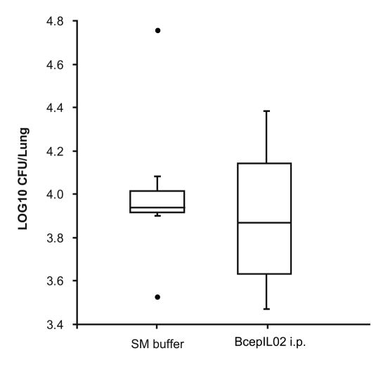
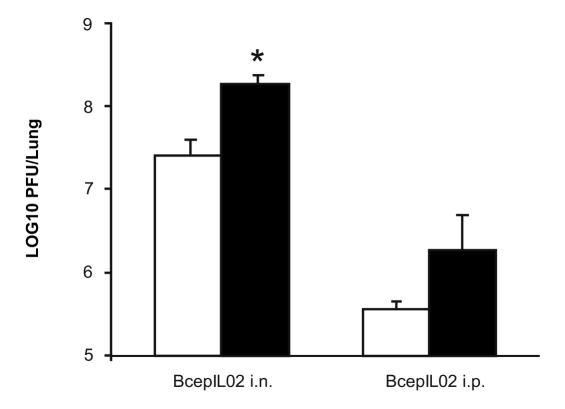
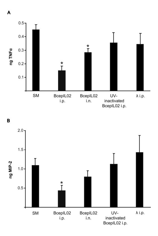
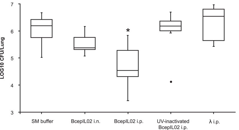
The therapeutic potential of bacteriophages (phages) in a mouse model of acute Burkholderia cenocepacia pulmonary infection was assessed. Phage treatment was administered by either intranasal inhalation or intraperitoneal injection. Bacterial density, macrophage inflammatory protein 2 (MIP-2), and tumor necrosis factor alpha (TNF-alpha) levels were significantly reduced in lungs of mice treated with intraperitoneal phages (P < .05). No significant differences in lung bacterial density or MIP-2 levels were found between untreated mice and mice treated with intranasal phages, intraperitoneal ultraviolet-inactivated phages, or intraperitoneal lambda phage control mice. Mock-infected mice treated with phage showed no significant increase in lung MIP-2 or TNF-alpha levels compared with mock-infected/mock-treated mice. We have demonstrated the efficacy of phage therapy in an acute B. cenocepacia lung infection model. Systemic phage administration was more effective than inhalational administration, suggesting that circulating phages have better access to bacteria in lungs than do topical phages.


Conflict of interest statement

Potential conflicts of interest: None of the authors has a commercial or other association that might pose a conflict of interest

Figures

FIG 1
FIG 1
Effect of phage treatment on bacterial density. Density of bacteria in lung was determined in mice that were infected with B. cenocepacia AU0728 and treated 24 h later with i.n. inhalation (n=6) or i.p. injection (n=6) of BcepIL02. Infected control mice were mock-treated with i.n. (n=5) or i.p. (n=11) SM buffer without phage (since there was no difference between these two groups, results were combined). Other control mice were treated with i.p. injection of UV-inactivated BcepIL02 (n=6), or i.p. injection of λ phage (n=8). The horizontal line in each box is the median CFU per lung for the group, calculated from two or more independent experiments, each performed in triplicate or quadruplicate; the boxes indicate the interquartile range of CFU per lung, and the whiskers indicate the range of CFU per lung for the group. ● = extreme outlier, * P < 0.05 (Mann-Whitney) for difference between BcepIL02 i.p. treated mice versus SM buffer mock-treated control mice.
FIG 2
FIG 2
Recovery of B. cenocepacia K56-2 from lungs 48 h after treatment. Density of bacteria was determined in mice that were infected with B. cenocepacia K56-2 and mock-treated 24 h later with SM buffer (n=7) or treated with i.p. injection of BcepIL02 (n=6). The horizontal line in each box is the median CFU per lung for the group, calculated from two independent experiments, each performed in triplicate or quadruplicate; the boxes indicate the interquartile range of CFU per lung, and the whiskers indicate the range of CFU per lung for the group ● = extreme outlier.
FIG 3
FIG 3
Recovery of phage from treated mice. Titer of BcepIL02 was determined in mice lungs that were infected with B. cenocepacia AU0728 (black bars, n=6) or mock-infected with SM buffer (white bars, n=6) and treated 24 h later with either i.n. inhalation or i.p. injection of BcepIL02. Data are mean + SEM PFU per lung 48 h after phage treatment, calculated from two independent experiments, each performed in triplicate. * P < 0.05 (Games-Howell) for difference between i.n BcepIL02 treatment of AU0728 infected versus mock-infected mice.
FIG 4
FIG 4
TNFα and MIP-2 levels in lungs after phage treatment. Levels of TNFα (A) and MIP-2 (B) were measured in mice lungs that were infected with AU0728. Twenty-four h after infection, mice were mock-treated with SM buffer (n=16), or treated with BcepIL02 by i.p. injection (n=6) or i.n. inhalation (n=6). Other control mice were treated via i.p. injection with UV-inactivated phage BcepIL02 (n=6) or λ phage (n=8). Data are mean + SEM ng TNFα or MIP-2 per lung, calculated from two or more independent experiments, each performed in triplicate or quadruplicate. * P < 0.05 (Games-Howell) for differences between treatments indicated and mock-treated SM buffer controls.
FIG 5
FIG 5
Immunofluorescent localization of B. cenocepacia AU0728. C57/BL6 mice were infected with B. cenocepacia AU0728. Lungs were removed 24 h (A) or 72 h (B) later, fixed, sectioned, stained for bacteria (red) and counterstained with DAPI (blue). A, Bacteria localized primarily in lung parenchyma with few observed in airway lumen. B, By 72 h after infection, bacteria formed microcolonies in areas of consolidated lung parenchyma. (A, B, bar = 20 μm).
FIG 6
FIG 6
Immunofluorescent localization of B. cenocepacia AU0728 and phage BcepIL02 in mouse lungs. C57BL/6 mice were infected with B. cenocepacia AU0728 and treated 24 h later by i.n. inhalation (A, B) or i.p. injection (C, D) of BcepIL02. Lungs were removed 48 h later, fixed, sectioned, and stained for phage (A-D; green) and bacteria (B, C, D; red). Phage administered via i.n. inhalation primarily localized in alveolar macrophages (A, B). B, Arrows indicate localization of degraded bacteria inside macrophages. Phage administered via i.p. injection were found primarily in perivascular areas (C) and alveolar septa, where they could be observed co-localized with degraded bacteria (D). (A, bar = 50 μm; B, C, bar = 20 μm; D, bar = 10 μm).

References

    1. Greenberg DE, Goldberg JB, Stock F, et al. Recurrent Burkholderia infection in patients with chronic granulomatous disease: 11-year experience at a large referral center. Clin Infect Dis. In Press. - PMC - PubMed
    1. LiPuma JJ. Update on the Burkholderia cepacia complex. Curr Opin Pulm Med. 2005;11:528–33. - PubMed
    1. Courtney JM, Dunbar KE, McDowell A, et al. Clinical outcome of Burkholderia cepacia complex infection in cystic fibrosis adults. J Cyst Fibros. 2004;3:93–8. - PubMed
    1. Isles A, Maclusky I, Corey M, et al. Pseudomonas cepacia infection in cystic fibrosis: An emerging problem. J Pediatr. 1984;104:206–10. - PubMed
    1. Reik R, Spilker T, LiPuma JJ. Distribution of Burkholderia cepacia complex species among isolates recovered from persons with or without cystic fibrosis. J Clin Microbiol. 2005;43:2926–8. - PMC - PubMed

Publication types