Review
. 2010 Aug 1;13(3):279-347.
doi: 10.1089/ars.2009.2788.

Potential therapeutic benefits of strategies directed to mitochondria

Affiliations
Review

Potential therapeutic benefits of strategies directed to mitochondria

Amadou K S Camara et al. Antioxid Redox Signal. .

Abstract

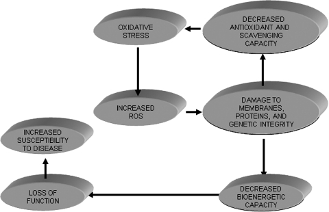
The mitochondrion is the most important organelle in determining continued cell survival and cell death. Mitochondrial dysfunction leads to many human maladies, including cardiovascular diseases, neurodegenerative disease, and cancer. These mitochondria-related pathologies range from early infancy to senescence. The central premise of this review is that if mitochondrial abnormalities contribute to the pathological state, alleviating the mitochondrial dysfunction would contribute to attenuating the severity or progression of the disease. Therefore, this review will examine the role of mitochondria in the etiology and progression of several diseases and explore potential therapeutic benefits of targeting mitochondria in mitigating the disease processes. Indeed, recent advances in mitochondrial biology have led to selective targeting of drugs designed to modulate and manipulate mitochondrial function and genomics for therapeutic benefit. These approaches to treat mitochondrial dysfunction rationally could lead to selective protection of cells in different tissues and various disease states. However, most of these approaches are in their infancy.


Figures

FIG. 1.
FIG. 1.
Basic mitochondrial structure and function. The figure shows the basic structural components of the five ETC complexes (I, II, III, IV, and V) as well as cytochrome c (Cyc), the flow of electrons through the complexes, and the generation of ATP. Fatty acid oxidation (FAO) and TCA cycle generate NADH and FADH2 needed to energize mitochondria and establish mitochondrial membrane potential (ΔΨm; −180 to −200 mV). ΔΨm is also modulated by uncoupling proteins (UCP). Phosphate carriers, including the adenine nucleotide translocase (ANT), regulate mitochondrial matrix phosphate levels. Substrate uptake is mediated through inner mitochondrial membrane (IMM) proteins [e.g., carnitine palmitoyl transferase (CPT) and pyruvate dehydrogenase (PDH)]. Mitochondrial DNA (mtDNA) encodes mitochondrial-specific proteins and cytosolic proteins produced by nuclear DNA (n) are translocated to mitochondria through the translocator of the outer membrane (TOM) and inner mitochondrial membrane (TIM); Ca2+ is taken up through the calcium uniporter (CaU). The mitochondrial Ca2+ level is dependent on the level of Ca2+ within the microdomain with the endoplasmic reticulum (ER). This basic function of mitochondria and its interaction with the nucleus and ER is the basis for understanding the role of the organelle in myriad of mitochondria-related diseases. Reproduced and modified from Wall et al. (604).
FIG. 2.
FIG. 2.
Putative mitochondrial permeability transition pore (mPTP) proteins (cylinders) in the outer (OMM) and inner (IMM) mitochondrial membrane in normal physiological and pathophysiological conditions. The constituents within the OMM include the VDAC, PBR, and other translocated proteins such as hexokinase (HK) and the Bcl family of proteins, Bax and Bak. The IMM proteins include ANT and CK; Cyclophilin D migrates to the IMM from the matrix. Initiators of apoptosis include cytochrome c and AIF. Small solutes include NAD+ and ADP. (For interpretation of the references to color in this figure legend, the reader is referred to the web version of this article at www.liebertonline.com/ars).
FIG. 3.
FIG. 3.
Some components of the mitochondrial ETC, OXPHOS, the TCA cycle, and various cation transporters and exchangers in IMM. Some aspects of the transporters and exchangers have been characterized computationally and pharmacologically. Protein components of the exchangers/transporters have yet to be fully characterized. Reproduced with permission and modified from Beard (47) and Dash and Beard (148).
FIG. 4.
FIG. 4.
Mitochondrial [Ca2+] before (baseline) and at 60 min reperfusion in isolated, constant pressure perfused hearts. (A) Time course of changes in systolic-diastolic (developed) left ventricular pressure (LVP) before during and after ischemia. (B) Hearts were perfused briefly for 10 min before and after ischemia (Bar line) with buffer (control), ruthenium red (RuR), an inhibitor of mitochondrial Ca2+ uptake, or amobarbital (AMO), a respiratory complex I inhibitor. Note that RuR, and to a greater extent AMO, improved function and reduced m[Ca2+] after ischemia. All data are expressed as means ± s.e.m. and statistical differences (between groups and within groups) were determined by two-way ANOVA. Differences between means were considered significant when p < 0.05 (two-tailed). If F tests were significant, appropriate post hoc analyses (Student–Newmann–Keuls or Duncan) were used to compare means. *p < 0.05 treatment vs. baseline/time control; †p < 0.05 pH 7.4 or pH 8.0 + RuR vs. other treatments; $p < 0.05 pH 7.4 alone vs. pH 8 alone; #p < 0.05 pH 7.4 + Amo vs. other treatments. Reproduced with permission of Aldakkak et al. (11).
FIG. 5.
FIG. 5.
Greater cyclosporine A (CsA) concentrations are required to increase Ca2+ uptake capacity in synaptic versus nonsynaptic mitochondrial fractions. Isolated synaptic or nonsynaptic mitochondria, in the presence or absence of 1 or 5 μM CsA, were placed in a constantly stirred, temperature-controlled, cuvette. CaG5N fluorescence, an indicator for Ca2+, was monitored continuously. Malate and pyruvate (M/P) and ADP were provided. Oligomycin (O), a complex V inhibitor, was added to ensure that the mitochondria were at maximal ΔΨm. The sharp rise in CaG5N fluorescence signifies mPTP opening and release of Ca2+ from mitochondria into the surrounding buffer. (A) In the absence of CsA, synaptic mitochondria sequestered much less Ca2+ than (B) nonsynaptic mitochondria. Arrows indicate the onset (On) and termination (Off ) of CaCl2 infusion. (C) Summary of quantitative estimates of Ca2+ uptake when Ca2+ was infused before mPTP opening. 1 μM CsA significantly increased Ca2+ uptake capacity of nonsynaptic mitochondria before mPTP opening but did not influence Ca2+ uptake capacity of synaptic mitochondria. CsA at 5 μM significantly increased Ca2+ uptake capacity of synaptic mitochondria compared with both 0 and 1 μM CsA. In contrast, 5 μM CsA did not further improve Ca2+ uptake capacity of nonsynaptic mitochondria compared to 1 μM CsA. In the presence of 5 μM CsA, Ca2+ uptake capacity of nonsynaptic mitochondria remained greater than that of synaptic mitochondria. *p < 0.05 indicates significant difference between groups (determined by one-way ANOVA and Scheffe's post hoc analysis). Reproduced with permission of Naga et al. (397).
FIG. 6.
FIG. 6.
A hypothetical model showing Bcl-2 and caspase regulation of Smac/DIABLO release from mitochondria. Cytochrome c and AIF released from mitochondria, as a result of mPTP opening or OMM permeabilization (Bad–Bax oligomerization), promote creation of the apoptosome, which triggers caspase-dependent feedback on mitochondria to release Smac/DIABLO. Smac/DIABLO in turn inhibits intrinsic IAPs, thereby neutralizing their caspase-inhibitory properties. This vicious cycle could continue to lead to more apoptosis, or it could be interrupted by the anti-apoptotic Bcl family of proteins, which by acting on the OMM reduce cytochrome c release and subsequently reduce Smac/DIABLO release and decrease apoptosis. The anti-caspase agent Z-VAD could also mitigate apoptosis by inhibiting the caspase-dependent mitochondrial attack. Reproduced with permission from Adrian et al. (5).
FIG. 7.
FIG. 7.
Differences in NADH/FAD (in arbitrary fluorescence units, afu) (A and B), ROS (in afu) (C), and LVP (in mmHg) (D), at baseline (BL), 30 min global ischemia, and at 5 and 60 min reperfusion with or without ranolazine, a putative respiratory complex I inhibitor. Ranolazine or vehicle (control) was infused for only 1 min just before ischemia. Ranolazine was not infused on reperfusion, but was present during ischemia. Note the improved redox state, reduced O2•− levels, and improved cardiac function after ranolazine treatment. All data are expressed as means ± s. e. m. and statistical differences (between groups and within groups) were determined by two-way ANOVA. Differences between means were considered significant when p < 0.05 (two-tailed). If F tests were significant, appropriate post hoc analyses (Student–Newman–Keuls or Duncan) were used to compare means. For p < 0.05: *Vehicle (control) vs. ranolazine. Preliminary data from Aldakkak et al. (8).
FIG. 8.
FIG. 8.
Mitochondrial O2•− generation (white stars) and antioxidant defense system (red stars). Mitochondria are primary consumers of O2 and are endowed with redox enzymes capable of transferring a single electron to O2 to generate O2•−. The sources of O2•− in mitochondria are discussed in detail in Section IV and the scavenging systems are presented in Section V. The sources of O2•− include MAO (monoamine oxidase) and cytochrome b5 reductase of the OMM; the ETC complexes and glycerol-3-phosphate dehydrogenase (GPDH) and pyruvate dehydrogenase (PDH) of the IMM; the TCA cycle enzymes, aconitase (Aco) and α-ketogluterate dehydrogenase (αKGDH). The transfer of electrons to O2 to generate O2•− is more likely when the redox carriers are fully reduced and ΔΨm is high. To minimize the level of O2•− within physiological range, mitochondria are replete with an elaborate antioxidant system to detoxify the O2•− generated by the reactions shown. In structurally intact mitochondria, a large scavenging capacity balances O2•− generation, and consequently, there is little net ROS production. The scavenging system consists of both nonenzymatic and enzymatic components. The nonenzymatic aspect includes cytochrome c (C), coenzyme Q10 (Q), and glutathione (GSH), and the enzymatic components include manganese superoxide dismutase (MnSOD), the so-called SOD2, catalase (Cat), glutathione peroxidase (GPX), glutathione reductase (GR), peroxiredoxins (PRX3/5), glutaredoxin (GRX2), thioredoxin (TRX2), and thioredoxin reductase (TrxR2). The regeneration of GSH (through GR) and reduced TRX2 (through TrxR2) depends on NADPH, which is derived from substrates or the membrane potential (through nicotinamide nucleotide transhydrogenase, TH). The antioxidant is also tied to the redox and energetic state of the mitochondrion (GSSG, glutathione disulphide, o, oxidized state; r, reduced state). The interplay between these redox systems (O2•− generation and scavenging) is vital for normal cellular function. Reproduced and modified from Lin and Beal (345). (For interpretation of the references to color in this figure legend, the reader is referred to the web version of this article at www.liebertonline.com/ars).
FIG. 9.
FIG. 9.
Mitochondrial ROS and RNS production and targets of oxidative and nitrosative damage on mitochondrial proteins. Mitochondrial NO activity most likely arises from extramitochondrial NOS. Mitochondria-derived NOS (mNOS) may also play a role, but this remains unresolved. The ONOO (NO + O2•−) arising from the extramitochondrial sources or formed intramitochondrially undergoes reactions in the different mitochondrial compartments and small amounts may escape to the cytosol. As shown, ONOO- targets several mitochondrial proteins important for normal physiological activity of the organelle. These include the ETC complexes, TCA cycle enzymes, and the scavenging system. The actions of RNS on these proteins could lead to mPTP opening and release of cytochrome c, or direct modulation of VDAC (voltage-dependent anion channel) and ANT (adenine nucleotide translocase) to release AIF (apoptosis inducing factor) and cytochrome c. S-nitrosation (SNO) play some regulatory role while nitration (NO2) reactions appear to be more permanent modifications and strictly linked to oxidative damage (Sox). Reproduced and modified from Radi et al. (461). (For interpretation of the references to color in this figure legend, the reader is referred to the web version of this article at www.liebertonline.com/ars).
FIG. 10.
FIG. 10.
Developed (systolic–diastolic) left ventricular pressure (LVP) in two groups of hearts exposed to either brief ischemic pulses (IPC), or to IPC bracketed by MnTBAP, an SOD mimetic. Developed LVP returned to pre-ischemia values between the IPC pulses and after the 2nd pulse (before index 30 min ischemia). On reperfusion after index ischemia, the IPC group had a better return of developed LVP than the ischemia control group, the IPC + MnTBAP group, or the MnTBAP group. Thus, ROS scavenging blocked the protection afforded by IPC. Statistical markings are shown in the figure after ANOVA and Student–Newman–Keuls tests of means from the different groups. Reproduced with permission of Kevin et al. (289).
FIG. 11.
FIG. 11.
Cardiac mitochondrial metabolism of different substrates in the normal state and in the pathological state (ischemia). In normal cardiomyocytes, cellular metabolism derives mostly from fatty acid metabolism. The transport of fatty acyl-CoA into mitochondria is accomplished via CPT 1. Once inside mitochondria, the fatty-CoA is a substrate for β-oxidation. During ischemia, substrate utilization is derived mostly from glucose and is less dependent on fatty acid metabolism. Ca2+ uptake through the Ca2+ uniporter (CaU) is thought to regulate TCA cycle enzyme activity. In the ischemic condition, Ca2+ uptake may occur via mPTP opening and other nonphysiological means (e.g., OMM permeabilization). Reproduced with permission and modified from Stark and Roden (540).
FIG. 12.
FIG. 12.
Summary of mitochondrial-targeted intervention and their therapeutic potential. See text for a more detailed description. Reproduced with permission and largely modified from Dykens et al. (179). (For interpretation of the references to color in this figure legend, the reader is referred to the web version of this article at www.liebertonline.com/ars).
FIG. 13.
FIG. 13.
Effect of SS-peptides on ischemia-induced GSH changes (A) and of S31 peptide on ischemia-induced infarct volume (Inf Vol) in C57BL/6 mice (B). (A) Mice were subjected to 30 min middle cerebral artery occlusion (MCAO) and treated with saline (Veh), SS31, or SS20 peptides immediately after reperfusion. Mice were sacrificed at 6 h post-ischemia. Values are expressed as GSH percent depletion in ipsilateral (Ipsil) compared with contralateral (Contral) cerebral hemispheres. Note that a difference was observed in %GSH depletion only in the SS31-treated cerebral cortex. (B) Mice were subjected to 30 min of MCAO and treated with saline/vehicle (Veh) or two different doses of SS31 immediately after reperfusion and at 6, 24, and 48 h reperfusion. Infarct volumes were estimated at 72 h post-ischemia from 12 serial sections (600 μm apart) per animal. SS31 reduced infarct size. Error bars indicate S.D. *p < 0.05 vs. Veh group, one-way ANOVA with post-hoc Newman–Keuls test. Reproduced with permission from Cho et al. (123).
FIG. 14.
FIG. 14.
Effect of anesthetic postconditioning (APoC) on mitochondrial pH (pHm) in SNARF-1 loaded myocytes. During hypoxia, pHm decreased as evidenced by a decrease in the SNARF-1 fluorescence ratio. Mitochondrial pH recovered immediately on reoxygenation. Treatment of cells with isoflurane at the beginning of reoxygenation delayed recovery of pHm, providing a more acidic matrix during early reoxygenation compared to the control group. Data are means ± S.D. Preliminary evidence provided by Pravdic et al. (456).
FIG. 15.
FIG. 15.
Ventricular infarct size expressed as % of total ventricular weight of guinea pig hearts at 120 min of reperfusion. Paxilline was administered for 10 min immediately after ischemia. Paxilline-treated hearts had larger infarcts than control hearts (A). Paxilline-treated hearts had less mitochondrial Ca2+ overload and a more reduced redox state (greater NADH) than the control (B). The Student's t-test was used to compare the means of the untreated group (control) vs. the treated group (paxilline). *p < 0.05 paxilline vs. control. Preliminary evidence provided by Varadarajan et al. (593).
FIG. 16.
FIG. 16.
ROS as a byproduct of oxidative stress and an essential component of some cellular functions and cell death. ROS are an integral component of the aging process, in which oxidative stress within mitochondria slowly degrades mitochondrial proteins, including the ETC complexes and matrix scavenging proteins. These alterations result in a self-perpetuating cycle of damage that eventually can lead to a decline in bioenergetic capacity and ultimately a compromise in organ system functional reserve. These factors predispose to increased probability and susceptibility damage. Reproduced with permission and modified from Muravchick et al. (391).

Similar articles

Cited by

References

    1. Abete P. Testa G. Ferrara N. De Santis D. Capaccio P. Viati L. Calabrese C. Cacciatore F. Longobardi G. Condorelli M. Napoli C. Rengo F. Cardioprotective effect of ischemic preconditioning is preserved in food-restricted senescent rats. Am J Physiol Heart Circ Physiol. 2002;282:H1978–H1987. - PubMed
    1. Abu–Hamad S. Zaid H. Israelson A. Nahon E. Shoshan–Barmatz V. Hexokinase-I protection against apoptotic cell death is mediated via interaction with the voltage-dependent anion channel-1: Mapping the site of binding. J Biol Chem. 2008;283:13482–13490. - PubMed
    1. Adams JM. Cory S. The Bcl-2 protein family: Arbiters of cell survival. Science. 1998;281:1322–1326. - PubMed
    1. Adams JM. Cory S. Bcl-2-regulated apoptosis: Mechanism and therapeutic potential. Curr Opin Immunol. 2007;19:488–496. - PMC - PubMed
    1. Adrain C. Creagh EM. Martin SJ. Apoptosis-associated release of Smac/DIABLO from mitochondria requires active caspases and is blocked by Bcl-2. EMBO J. 2001;20:6627–6636. - PMC - PubMed

Publication types

LinkOut - more resources