. 2010 Feb 24;426(3):293-306.
doi: 10.1042/BJ20091459.

Post-transcriptional regulation of MEK-1 by polyamines through the RNA-binding protein HuR modulating intestinal epithelial apoptosis

Affiliations

Post-transcriptional regulation of MEK-1 by polyamines through the RNA-binding protein HuR modulating intestinal epithelial apoptosis

Peng-Yuan Wang et al. Biochem J. .

Abstract

MEK-1 [MAPK (mitogen-activated protein kinase) kinase-1] is an important signal transducing enzyme that is implicated in many aspects of cellular functions. In the present paper, we report that cellular polyamines regulate MEK-1 expression at the post-transcriptional level through the RNA-binding protein HuR (Hu-antigen R) in IECs (intestinal epithelial cells). Decreasing the levels of cellular polyamines by inhibiting ODC (ornithine decarboxylase) stabilized MEK-1 mRNA and promoted its translation through enhancement of the interaction between HuR and the 3'-untranslated region of MEK-1 mRNA, whereas increasing polyamine levels by ectopic ODC overexpression destabilized the MEK-1 transcript and repressed its translation by reducing the abundance of HuR-MEK-1 mRNA complex; neither intervention changed MEK-1 gene transcription via its promoter. HuR silencing rendered the MEK-1 mRNA unstable and inhibited its translation, thus preventing increases in MEK-1 mRNA and protein in polyamine-deficient cells. Conversely, HuR overexpression increased MEK-1 mRNA stability and promoted its translation. Inhibition of MEK-1 expression by MEK-1 silencing or HuR silencing prevented the increased resistance of polyamine-deficient cells to apoptosis. Moreover, HuR overexpression did not protect against apoptosis if MEK-1 expression was silenced. These results indicate that polyamines destabilize the MEK-1 mRNA and repress its translation by inhibiting the association between HuR and the MEK-1 transcript. Our findings indicate that MEK-1 is a key effector of the HuR-elicited anti-apoptotic programme in IECs.


Figures
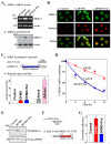
Fig. 1
Fig. 1
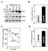
Polyamine depletion increases MEK-1 expression. (A) Levels of MEK-1 mRNA and protein in cells exposed to DFMO (5 mM) alone or DFMO plus putrescine (Put, 10 μM) for 6 days: a) changes in MEK-1 mRNA as measured by RT-PCR analysis; and b) representative immunoblots of MEK-1 protein by Western analysis. The first-strand cDNAs, synthesized from total cellular RNA, were amplified with the specific sense and antisense primers, and PCR-amplified products displayed in agarose gel for MEK-1 (~450 bp) and β-actin (~244 bp). To measure levels of MEK-1 protein, 20 μg of total proteins were applied to each lane, and immunoblots were hybridized with the antibody specific for MEK-1 (~45 kDa). Actin (~42 kDa) immunoblotting was performed as an internal control for equal loading. (B) Cellular distribution of MEK-1: a) control; b) DFMO-treated cells; and c) cells treated with DFMO plus Put. Cells were permeabilized and incubated with the anti-MEK-1 antibody and then with anti-IgG conjugated with Alexa Fluor. Nuclei were stained with the TO-PRO3. Green, MEK-1 signals; red, nuclei. Original magnification, ×1,000. (C) Levels of MEK-1-promoter activity in cells described in A: a) schematic MEK-1-promoter luciferase (Luc) reporter construct (pMEK1-luc); b) levels of luciferase reporter activity after polyamine depletion. After cells were treated with DFMO or DFMO plus Put for 4 days, they were transfected with the pMEK1-luc or control vector (pGL3) using LipofectAMINE technique; luciferase activity was examined 48 h after transfection. In a separate study, cells were cotransfected with the pMEK1 and the c-jun expression vector (Adc-jun) or control vector (Adnull), and luciferase activity was assayed 48 h thereafter. Data were normalized by Renilla-driven luciferase activity and expressed as means ± SE of data from 3 separate experiments. * P < 0.05 compared with controls and cells infected with Adnull. (D) Half-life of MEK-1 mRNA in cells described in A. After cells were incubated with actinomycin D for the indicated times, total cellular RNA was isolated, and the levels of remaining MEK-1 and GAPDH mRNAs were measured by Q-PCR analysis. Values are the means ± SE from triplicate samples. (E) Newly translated MEK-1 protein in cells described in A. MEK-1 translation was measured by incubating cells with L-[35S]methionine and L-[35S]cysteine for 20 min, followed by immunoprecipitation by using anti-MEK-1 antibody, resolving immunoprecipitated samples by SDS/PAGE, and transferring for visualization of signals by using a PhosphorImager. The translation of housekeeping control GAPDH was measured similarly. (F) Schematic of plasmids: a) control (pGL3-Luc); b) chimeric firefly luciferease (Luc)-MEK-1 3′UTR (pGL3-Luc-MEK1ARE). (G) Changes in MEK-1 translation efficiency as measured by using pGL3-Luc-MEK1ARE reporter assays in cells described in A. The pGL3-Luc-MEK1ARE or pGL3-Luc (negative control) was cotransfected with a Renilla luciferase reporter. Twenty-four hours later, firefly and Renilla luciferase activities were assayed. Luciferase values were normalized to the mRNA levels to obtain translation efficiencies and expressed as means ± SE of data from 3 separate experiments. * p < 0.05 compared with controls and cells treated with DFMO plus Put.
Fig. 2
Fig. 2
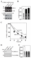
Increasing cellular polyamines represses MEK-1 expression. (A) Changes in MEK-1 mRNA and protein in clonal (C) populations of ODC-IEC cells (ODC) and control cells (C-vector). IEC-6 cells were infected with either the retroviral vector containing the sequence encoding mouse ODC cDNA or control retroviral vector lacking ODC cDNA. Clones resistant to the selection medium containing 0.6 mg/ml G418 were isolated and screened for ODC expression. The levels of MEK-1 mRNA and protein were assessed by RT-PCR analysis and Western immunoblotting, respectively. (B) Levels of MEK-1-promoter activity in cells described in A. Luciferase activity was examined 48 h after transfection with pMEK1-luc or the control vector (pGL3). Data were normalized by Renilla-driven luciferase activity and expressed as means ± SE of data from 3 separate experiments. (C) Half-life of MEK-1 mRNA in cells described in A. After cells were incubated with actinomycin D for the indicated times, total cellular RNA was isolated, and the levels of remaining MEK-1 and GAPDH mRNAs were measured by Q-PCR analysis. Values are the means ± SE from triplicate samples. (D) Newly synthesized MEK-1 protein in cells described in A. After cells were incubated with L-[35S]methionine and L-[35S]cysteine for 20 min, cell lysates were prepared and immunoprecipitated by using anti-MEK-1 antibody, resolved by SDS/PAGE, and transferred for visualization of signals by using a PhosphorImager. The translation of housekeeping control GAPDH was measured similarly. (E) Changes in MEK-1 translation efficiency as measured by using pGL3-Luc-MEK1ARE reporter assays in cells described in A. The pGL3-Luc-MEK1ARE or pGL3-Luc (negative control) was cotransfected with a Renilla luciferase reporter, and firefly and Renilla luciferase activities were assayed 24 h thereafter. Luciferase values were normalized to the mRNA levels to obtain translation efficiencies and expressed as means ± SE of data from 3 separate experiments. * p < 0.05 compared with controls.
Fig. 3
Fig. 3
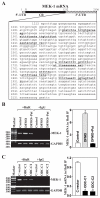
Changes in HuR-binding to the MEK-1 mRNA after altering the levels of cellular polyamines. (A) Schematic representative of the MEK-1 mRNA and the predicted hits of the HuR signature motif in its 3′-UTR as indicated by underline. (B) Levels of association of endogenous HuR with endogenous MEK-1 mRNA after polyamine depletion. IEC-6 cells were exposed to DFMO alone or DFMO plus putrescine for 6 days. After immunoprecipitation (IP) of RNA-protein complexes from cell lysates using either anti-HuR antibody (Ab) or control IgG1, RNA was isolated and used in RT reactions. Left panel: representative RT-PCR products visualized in ethidium bromide-stained agarose gels; low-level amplification of GAPDH (housekeeping mRNA, which is not HuR targets) served as negative controls. Right panel: fold differences in MEK-1 transcript abundance in HuR IP compared with IgG IP, as measured by Q-PCR analysis. Values were means ± SE from triplicate samples. * p < 0.05 compared with controls and cells treated with DFMO plus Put. (C) Changes in levels of MEK-1 mRNA in HuR IP from clonal (C) populations of ODC-IEC cells (ODC) and control cells (C-vector). Left panel: representative RT-PCR products of MEK-1 and GAPDH mRNAs in HuR or IgG1 IP. Right panel: fold differences in MEK-1 mRNA in HuR IP compared with IgG IP as measured by Q-PCR analysis. Values were means ± SE from triplicate samples. * p < 0.05 compared with controls.
Fig. 4
Fig. 4
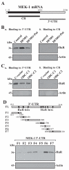
HuR-binding to the 3′-UTR and coding region (CR) of MEK-1 mRNA in cells described in Fig. 3. (A) Schematic representation of the MEK-1 biotinylated transcripts (CR and 3′-UTR) used in this study. (B) Representative HuR immunoblots using the pulldown materials by different fractions of MEK-1 mRNA after polyamine depletion: a) HuR-binding to 3′-UTR; and b) HuR-binding to CR. Cytoplasmic lysates prepared from control cells and cells exposed to DFMO alone or DFMO plus Put for 6 days were incubated with 6 μg of biotinylated MEK-1 3′-UTR or CR for 30 min at 25 °C, and the resulting RNP complexes were pulled down by using streptavidin-coated beads. The presence of HuR in the pull-down material was assayed by Western blotting. β-actin in the pull-down material was also examined and served as a negative control. (C) Representative HuR immunoblots using the pulldown materials in clonal (C) populations of ODC-IEC cells (ODC) and control cells (C-vector): a) HuR-binding to 3′-UTR; and b) HuR-binding to CR. (D) Representative HuR immunoblots in the material pulled down by different biotinylated fractions of the MEK-1 mRNA 3′-UTR. Top panel, schematic representation of the MEK-1 3′-UTR biotinylated transcripts used in this study. Three experiments were performed that showed similar results.
Fig. 5
Fig. 5
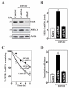
Effect of HuR silencing on MEK-1 mRNA stability and its translation in polyamine-deficient cells. (A) Representative HuR and MEK-1 immunoblots. After cells were cultured in the presence of DFMO for 4 days, they were transfected with either siRNA targeting the HuR mRNA coding region (siHuR) or control siRNA (C-siRNA), and whole-cell lysates were harvested 48 h thereafter. The levels of HuR and MEK-1 proteins were measured by Western blot anaylsis, and equal loading was monitored by β-actin immunoblotting. (B) Levels of MEK-1 mRNA in cells that were processed as described in panel A. Total RNA from each group was harvested, and levels of MEK-1 mRNA were measured by RT + Q-PCR analysis. The data were normalized to the amount of GAPDH mRNA and the values represented as the means ± SE of data from triplicate experiments. * P < 0.05 compared with controls and cells transfected with C-siRNA. (C) Half-life of the MEK-1 mRNA in cells that were transfected and treated as described in A. Total cellular RNA was isolated at the indicated times after administration of actinomycin D, and the remaining levels of MEK-1 and GAPDH mRNAs were measured by RT + Q-PCR analysis. Values are means ± SE from triplicate samples. (D) Changes in MEK-1 translation efficiency as measured by using pGL3-Luc-MEK1ARE reporter assays in cells described in A. The pGL3-Luc-MEK1ARE or pGL3-Luc (negative control) was cotransfected with a Renilla luciferase reporter, and firefly and Renilla luciferase activities were assayed 24 h thereafter. Luciferase values were normalized to the mRNA levels to obtain translation efficiencies and expressed as means ± SE of data from 3 separate experiments. * p < 0.05 compared with control cells and DFMO-treated cells transfected with siHuR.
Fig. 6
Fig. 6
Changes in MEK-1 mRNA stability and translational efficiency after ectopic HuR overexpression. (A) Representative immunoblots of HuR and MEK-1 proteins after ectopic HuR expression. Cells were infected with the recombinant adenoviral vector encoding HuR cDNA (AdHuR, prepared as explained in Materials and Methods) or adenoviral vector lacking HuR cDNA (Adnull) at a multiplicity of infection of 50-200 plaque-forming units (pfu)/cell; the expression of HuR and MEK-1 proteins was analyzed 48 h after the infection. (B) Levels of MEK-1 mRNA as measured by RT-qPCR analysis in cells infected with AdHuR or Adnull at the concentration of 100 pfu/cell for 48 h. Data were normalized to amount of GAPDH mRNA, and values are means ± SE of data from triplicate experiments. * P < 0.05 compared with cells infected with Adnull. (C) Half-life of the MEK-1 mRNA as measured by RT-qPCR analysis by using actinomycin D in cells described in B. Values are the means ± SE from triplicate samples. (D) Changes in MEK-1 translation efficiency as measured by using pGL3-Luc-MEK1ARE reporter assays in cells that were processed as described in B. Twenty-four h after cells were transfected with the pGL3-Luc-MEK1ARE or pGL3-Luc (negative control), the levels of luciferase activity were examined and normalized to the mRNA levels to obtain translation efficiencies. Values were expressed as means ± SE of data from 3 separate experiments. * p < 0.05 compared with cells transfected with Adnull.
Fig. 7
Fig. 7
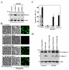
Effects of MEK-1 or HuR silencing on apoptosis in polyamine-deficient cells. (A) Cells were cultured with DFMO for 4 days and then transfected with either siRNA specifically targeting the MEK-1 mRNA coding region (siMEK-1), siHuR, or C-siRNA. The levels of MEK-1 protein were measured by Western blot analysis 48 h after transfection; equal loading was monitored by β-actin immunoblotting. (B) TNFα/CHX-induced apoptosis in cells treated as described in panel A: a) control cells; b) DFMO-treated cells transfected with C-siRNA; c) DFMO-treated cells transfected with siHuR; d) DFMO-treated cells transfected with siMEK-1. Apoptosis was measured by morphological analysis (middle) and ApoAlert annexin-V (A-V) staining (right) 4 h after treatment with TNFα/CHX. Original magnification, ×150. (C) Percentage of apoptotic cells in cultures processed as described in panel B. Values are the means ± SE from six samples. * p < 0.05 compared with cells that were not treated with TNFα/CHX. + p < 0.05 compared with DFMO-treated cells that were transfected with C-siRNA and then treated with TNFα/CHX for 4 h. (D) Representative immunoblots for procaspase-3 and caspase-3 in cells that were processed as described in panel B. Whole-cell lysates were harvested 4 h after treatment with TNFα/CHX, and the levels of procaspase-3 and caspase-3 were examined by Western blot analysis. Three experiments were performed that showed similar results.
Fig. 8
Fig. 8
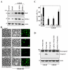
Effect of MEK-1 silencing on apoptotic sensitivity in cells overexpressing HuR. (A) Representative HuR and MEK-1 immunoblots. Cells were transfected with either siMEK-1 or C-siRNAfor 24 h and then infected with AdHuR or Adnull (100 pfu/cell). HuR and MEK-1 protein levels were examined by Western blot analysis 24 h after infection, and equal loading was monitored by β-actin immunoblotting. (B) TNFα/CHX-induced apoptosis in cells described A: a) cells infected with Adnull; b) cells infected with AdHuR; c) cells infected with AdHuR and transfected with C-siRNA; d) cells infected with AdHuR and transfected with siMEK-1. Apoptosis was measured by morphological analysis (middle) and ApoAlert annexin-V (A-V) staining (right) 4 h after treatment with TNFα/CHX. Original magnification, ×150. (C) Percentage of apoptotic cells as described in B. Values are means ± SE of data from six samples. * p < 0.05 compared with groups that were not treated with TNFα/CHX. + p < 0.05 compared with cells infected with Adnull and then treated with TNFα/CHX. (D) Representative immunoblots for procaspase-3 and caspase-3 in cells that were processed as described in B. Whole-cell lysates were harvested 4 h after treatment with TNFα/CHX, and the levels of procaspase-3 and caspase-3 proteins were examined by Western blot analysis. Three experiments were performed that showed similar results.

Similar articles

Cited by

References

    1. Radtke F, Clevers H. Self-renewal and cancer of the gut: two sides of a coin. Science. 2005;307:1904–1909. - PubMed
    1. Wang JY. Polyamines and mRNA stability in regulation of intestinal mucosal growth. Amino. Acids. 2005;33:241–252. - PubMed
    1. Orphanides G, Reinberg D. A unified theory of gene expression. Cell. 2002;108:439–451. - PubMed
    1. Li L, Liu L, Rao JN, Esmaili A, Strauch ED, Bass BL, Wang JY. JunD stabilization results in inhibition of normal intestinal epithelial cell growth through p21 after polyamine depletion. Gastroenterology. 2002;123:764–779. - PubMed
    1. Li L, Rao JN, Guo X, Liu L, Santora R, Bass BL, Wang JY. Polyamine depletion stabilizes p53 resulting in inhibition of normal intestinal epithelial cell proliferation. Am. J. Physiol. Cell. Physiol. 2001;281:C941–C953. - PubMed

Publication types

MeSH terms