. 2010 Jan;159(1):237-51.
doi: 10.1111/j.1476-5381.2009.00517.x. Epub 2009 Nov 27.

Role of the signal peptide in the synthesis and processing of the glucagon-like peptide-1 receptor

Affiliations

Role of the signal peptide in the synthesis and processing of the glucagon-like peptide-1 receptor

Y Huang et al. Br J Pharmacol. 2010 Jan.

Abstract

Background and purpose: The glucagon-like peptide-1 receptor (GLP-1R) belongs to Family B of the G protein-coupled receptor superfamily and is a target for treatment of type 2 diabetes. Family B G protein-coupled receptors contain a putative N-terminal signal peptide, but its role in receptor synthesis and trafficking are unclear. Further, the signal peptide is not cleaved in at least one family member.

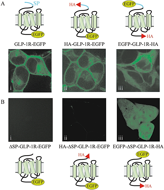
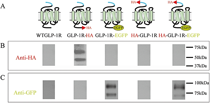
Experimental approach: We examined receptor glycosylation and the role of the signal peptide in GLP-1R synthesis and trafficking using constructs containing epitope tags at the N- and/or C-terminus and in which the signal peptide sequence was either present or absent.

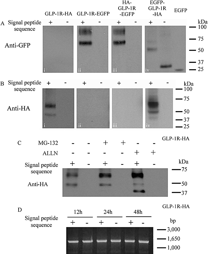
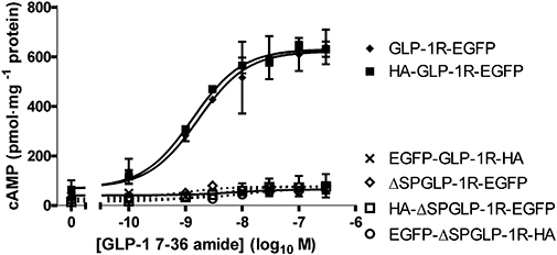
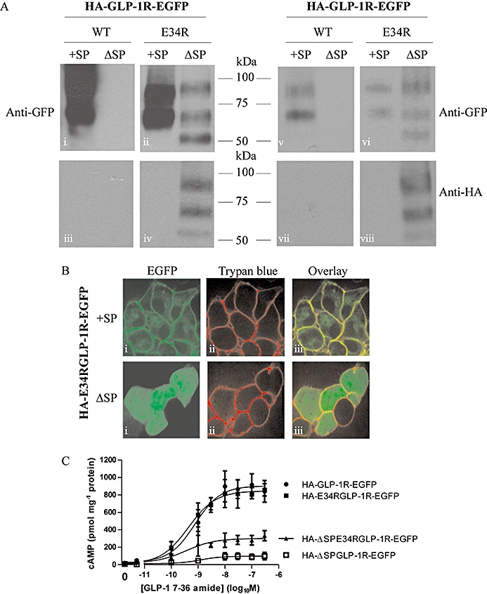
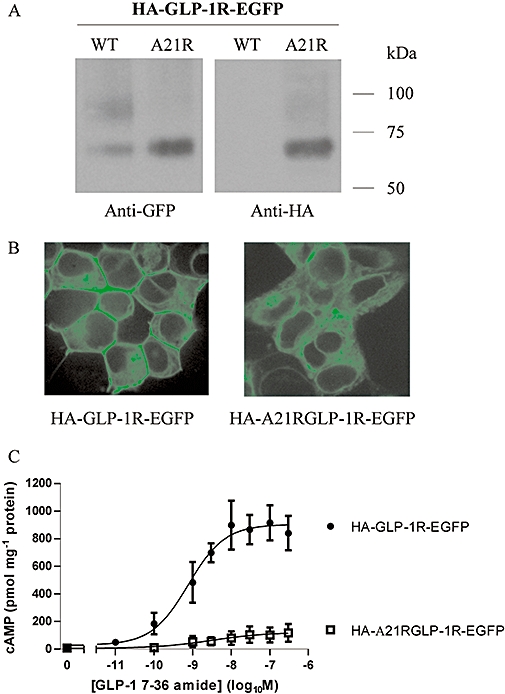
Key results: The signal peptide was absolutely required for GLP-1R synthesis but could be substituted to some extent by increasing positive charge in the N-terminal region of the receptor flanking the signal peptide. The signal peptide is cleaved during synthesis and processing of the receptor. An enhanced GFP-epitope tag at the N-terminus of the receptor permitted synthesis of the receptor but blocked signal peptide cleavage and prevented trafficking to the plasma membrane. Cleavage site mutation allowed synthesis of a full-length receptor, blocked signal peptide cleavage and caused retention within the endoplasmic reticulum.

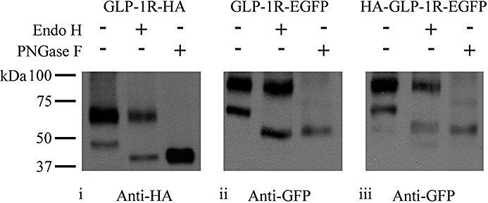
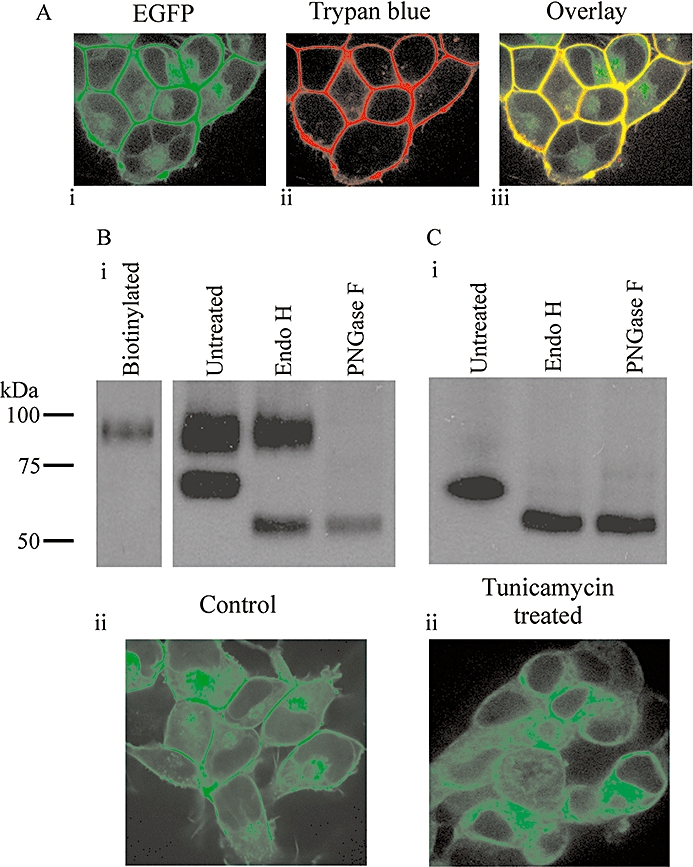
Conclusions and implications: Signal peptide cleavage was not essential for receptor synthesis but was obligatory for processing and trafficking of receptors to the plasma membrane. Further, the GLP-1R is subject to N-linked glycosylation and only the mature, fully glycosylated form of the receptor is present in the plasma membrane. Inhibition of glycosylation prevents processing and cell surface expression of the GLP-1R.


Figures

Figure 2
Figure 2
Confocal microscopy of cells transfected with EGFP-tagged glucagon-like peptide-1 receptors (GLP-1Rs) with or without the signal peptide sequence. Human embryonic kidney (HEK)-293 cells were transiently transfected with plasmids containing GLP-1R constructs that had an EGFP-epitope tag at either the C-terminus (GLP-1R-EGFP, HA-GLP-1R-EGFP) or N-terminus (EGFP-GLP-1R-HA) and that either contained the signal peptide sequence (A) or had it deleted [ΔSP; (B)]. After 30–48 h, live cells were examined by confocal microscopy. Images are representative of cells observed in five independent experiments.
Figure 1
Figure 1
Immunoblotting of cell lysates following transfection of cells with glucagon-like peptide-1 receptor (GLP-1R) constructs. Human embryonic kidney (HEK)-293 cells were transiently transfected with plasmids containing either the WTGLP-1R or various epitope-tagged constructs as indicated (A). After approximately 30 h, cell lysates were prepared and immunoblotted using either an anti-HA antibody (B) or an anti-GFP antibody (C). Data are representative of three independent experiments.
Figure 4
Figure 4
Functional coupling of glucagon-like peptide-1 receptor (GLP-1R) constructs. Human embryonic kidney (HEK)-293 cells were transiently transfected with plasmids containing either the WTGLP-1R or various epitope-tagged constructs. After 30 h, cells were challenged with GLP-1 7-36 amide at the concentrations indicated and cAMP levels determined after 10 min stimulation. Data are mean ± SEM, n= 3. pEC50 values are given in Table 2.
Figure 3
Figure 3
Immunoblotting of cell lysates following transfection of epitope-tagged receptors with or without the signal peptide sequence. Human embryonic kidney (HEK)-293 cells were transiently transfected with plasmids containing various epitope-tagged constructs that either contained the signal peptide sequence (+) or in which it had been deleted (−). After 24–30 h, cell lysates were prepared and immunoblotted using either an anti-HA (A) or an anti-GFP (B) antibody as appropriate. Additionally, groups of cells were transfected with the pEGFP-C1 plasmid to allow expression of EGFP alone. (C) Cells were transfected with either GLP-1R-HA or the signal peptide-deleted equivalent (ΔSP-GLP-1R-HA) and 24 h later treated for a further 6 h with either the proteasome and cathepsin K inhibitor, MG132 (10 µM) or the proteasome and calpain 1 inhibitor, ALLN (MG101; 261 µM/100 µg·mL−1). (D) Cells were transfected with either GLP-1R-HA or ΔSP-GLP-1R-HA and RT-PCR performed at 12, 24 and 48 h using identical forward and reverse primers designed to amplify the sequence representing the full-length receptor but excluding the signal peptide sequence. Data are representative of three independent experiments.
Figure 5
Figure 5
Functional coupling of glucagon-like peptide-1 receptor (GLP-1R) constructs with or without the signal peptide sequence. Human embryonic kidney (HEK)-293 cells transiently transfected with epitope-tagged GLP-1Rs either with or without the signal peptide sequence were assessed for coupling to the generation of cAMP. Cells were challenged with GLP-1 7-36 amide at the concentrations indicated and cAMP levels determined after 10 min stimulation. Data are mean ± SEM, n= 3. pEC50 values for those receptors having the signal peptide sequence and in which cAMP responses were measurable (GLP-1R-EGFP and HA-GLP-1R-EGFP) were similar to those in Table 2.
Figure 6
Figure 6
Increasing positive charge within the N-terminus of the receptor partially compensates for the lack of a signal peptide. Human embryonic kidney (HEK)-293 cells were transiently transfected with the glucagon-like peptide-1 receptor (GLP-1R) construct containing an N-terminal HA-tag and C-terminal EGFP tag either with (HA-GLP-1R-EGFP) or without (HA-ΔSP-GLP-1R-EGFP) the signal peptide. We also used similar constructs in which the glutamic acid residue at position 34 had been mutated to an arginine (HA-E34R-GLP-1R-EGFP and HA-ΔSP-E34R-GLP-1R-EGFP). After 24–30 h, cell lysates were prepared and immunoblotted using either an anti-HA or an anti-GFP antibody (A). In the left-hand panel, lanes were equally loaded, showing the relatively poor but nevertheless clear expression of the HA-ΔSP-E34R-GLP-1R-EGFP construct. In the right-hand panels, lanes for the constructs containing the signal peptide sequence (+SP) were loaded with fivefold less protein than lanes for the signal peptide-deleted constructs (ΔSP) to more clearly show the relative patterns of immunoreactive bands. Live cells were examined by confocal microscopy (B) or challenged with GLP-1 7-36 amide at the concentrations indicated and cAMP levels determined after 10 min stimulation (C). Immunoblots and confocal images are representative of three independent experiments while data for cAMP generation are mean ± SEM, n= 3–6. pEC50 values: HA-GLP-1R-EGFP, 9.12 ± 0.24; HA-ΔSP-GLP-1R-EGFP, 9.36 ± 0.15; HA-E34R-GLP-1R-EGFP, 8.74 ± 0.47; HA-ΔSP-E34R-GLP-1R-EGFP, 8.46 ± 0.62.
Figure 7
Figure 7
Mutation of the predicted signal peptide cleavage site blocks both cleavage and plasma membrane expression of the glucagon-like peptide-1 receptor (GLP-1R). Human embryonic kidney (HEK)-293 cells were transiently transfected with either HA-GLP-1R-EGFP or this construct containing an alanine to arginine mutation at position −3 relative to the predicted cleavage site (HA-A21R-GLP-1R-EGFP). After 24–30 h, cell lysates were prepared and immunoblotted using either an anti-HA or an anti-GFP antibody (A). Live cells were examined by confocal microscopy (B) or challenged with GLP-1 7-36 amide at the concentrations indicated and cAMP levels determined after 10 min stimulation (C). Immunoblots and confocal images are representative of three independent experiments while data for cAMP generation are mean ± SEM, n= 3. pEC50 value for HA-GLP-1R-EGFP, 9.09 ± 0.22.
Figure 8
Figure 8
Glycosylation of the glucagon-like peptide-1 receptor (GLP-1R). Human embryonic kidney (HEK)-293 cells were transiently transfected with plasmids containing epitope-tagged GLP-1Rs as indicated. After 48 h, cell lysates were prepared and either untreated or subjected to treatment with either Endo H or PNGase F as described in Methods. Lysates were then processed and immunoblotted using either an anti-HA (i) or anti-GFP (ii and iii) antibody as indicated. Data are representative of three independent experiments.
Figure 9
Figure 9
Influence of the glycosylation inhibitor, tunicamycin, on the glycosylation pattern and subcellular distribution of the glucagon-like peptide-1 receptor (GLP-1R). Human embryonic kidney (HEK)-293 cells with stable expression of the C-terminal, EGFP-tagged GLP-1R (GLP-1R-EGFP) were either untreated (A and B) or treated with tunicamycin (C) for 7.5 days as described in Methods. Cells were then examined by confocal microscopy (A, Bii and Cii), including following membrane staining with trypan blue (A) or lysates prepared and either untreated or subjected to treatment with either Endo H or PNGase F as described. Lysates were then processed and immunoblotted using an anti-GFP antibody (Bi and Ci). Alternatively, proteins at the plasma membrane of intact cells were biotinylated, captured using streptavidin and subject to immunoblotting using the anti-GFP antibody (Bi; ‘biotinylated’). Data are representative of six coverslips and three immunoblots.

Similar articles

Cited by

References

    1. Alexander SPH, Mathie A, Peters JA. Guide to receptors and channels (GRAC), 3rd edition (2008 revision) Br J Pharmacol. 2008;153(Suppl. 2):S1–S209. - PMC - PubMed
    1. Alken M, Rutz C, Köchl R, Donalies U, Oueslati M, Furkert J. The signal peptide of the rat corticotropin-releasing factor receptor 1 promotes receptor expression but is not essential for establishing a functional receptor. Biochem J. 2005;390:455–464. - PMC - PubMed
    1. Alken M, Schmidt A, Rutz C, Furkert J, Kleinau G, Rosenthal W, et al. The sequence after the signal peptide of the G protein-coupled endothelin B receptor is required for efficient translocon gating at the endoplasmic reticulum membrane. Mol Pharmacol. 2009;75:801–811. - PubMed
    1. Baggio LL, Drucker DJ. Biology of incretins: GLP-1 and GIP. Gastroenterology. 2007;132:2131–2157. - PubMed
    1. Bendtsen JD, Nielsen H, von Heijne G, Brunak S. Improved prediction of signal peptides: SignalP 3.0. J Mol Biol. 2004;340:783–795. - PubMed

Publication types