. 2010 Jan;159(1):222-36.
doi: 10.1111/j.1476-5381.2009.00516.x. Epub 2009 Nov 27.

Differential actions of urocortins on neurons of the myenteric division of the enteric nervous system in guinea pig distal colon

Affiliations

Differential actions of urocortins on neurons of the myenteric division of the enteric nervous system in guinea pig distal colon

Sumei Liu et al. Br J Pharmacol. 2010 Jan.

Abstract

Background and purpose: Urocortins (Ucns) 1, 2 and 3 are corticotropin-releasing factor (CRF)-related neuropeptides and may be involved in neural regulation of colonic motor functions. Nevertheless, details of the neural mechanism of action for Ucns have been unclear. We have, here, tested the hypothesis that Ucns act in the enteric nervous system (ENS) to influence colonic motor behaviour.

Experimental approach: We used intracellular recording with 'sharp' microelectrodes, followed by intraneuronal injection of biocytin, and immunohistochemical localization of CRF(1) and CRF(2) receptors in guinea pig colonic tissue.

Key results: Application of Ucn1 depolarized membrane potentials and elevated excitability in 58% of AH-type and 60% of S-type colonic myenteric neurons. In most of the neurons tested, depolarizing responses evoked by Ucn-1 were suppressed by the CRF(1) receptor antagonist NBI 27914, but were unaffected by the CRF(2) receptor antagonist antisauvagine-30. The selective CRF(2) receptor agonists, Ucn2 and Ucn3, evoked depolarizing responses in 12 and 8% of the AH-type myenteric neurons, respectively, and had no effect on S-type neurons. Antisauvagine-30, but not NBI 27914, suppressed these Ucn2- and Ucn3-evoked responses. Immunohistochemical staining identified CRF(1) as the predominant CRF receptor subtype expressed by ganglion cell somas, while CRF(2)-immunoreactive neuronal somas were sparse. Ucns did not affect excitatory synaptic transmission in the ENS.

Conclusions and implications: The results suggest that Ucns act as neuromodulators to influence myenteric neuronal excitability. The excitatory action of Ucn1 in myenteric neurons was primarily at CRF(1) receptors, and the excitatory action of Ucn2 and Ucn3 was at CRF(2) receptors.


Figures

Figure 1
Figure 1
Expression of CRF1 and CRF2 receptor immunoreactivity (IR) in the myenteric plexus of the guinea pig distal colon. (A1) CRF1 receptor IR was expressed in neuronal cell bodies within the myenteric plexus. (A2) Pre-absorption of the CRF1 receptor antibody with the blocking peptide (sc-12383p) eliminated specific staining. (A3) Western blot analysis demonstrated that the CRF1 receptor antibody recognized a protein band around 66 kDa in samples extracted from longitudinal muscle–myenteric plexus (LMMP) and hypothalamus. Pre-absorption with the blocking peptide against which the CRF1 receptor antibody was raised resulted in the loss of the immunoreactive band (control). (B1) CRF2 receptor IR was expressed predominantly in varicose nerve fibres in the myenteric plexus. CRF2-immunoreactive neuronal cell bodies were sparse. (B2) Omitting the primary antibody for CRF2 receptors eliminated the immunostaining. (B3) Western blot analysis demonstrated that the CRF2 receptor antibody recognized a protein band around 70 kDa in samples extracted from LMMP, cerebellum and hypothalamus. (C) CRF2 receptor IR was expressed in the guinea pig hypothalamus, which served as a positive control. LMMP, longitudinal muscle–myenteric plexus; Hypo, hypothalamus; C, cerebellum. Scale bars = 20 µm.
Figure 3
Figure 3
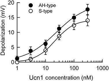
Concentration–response relationships for urocortin-1 (Ucn1)-evoked membrane depolarization in AH- and S-type neurons. Each data point represents six AH-type neurons and six S-type neurons. The EC50 value was 37.6 ± 9.8 nM for AH-type neurons, and 60.5 ± 18.9 nM for S-type neurons.
Figure 2
Figure 2
Depolarizing responses to urocortin-1 (Ucn1) were concentration dependent in both AH- and S-type colonic myenteric neurons. (A) Responses to Ucn1 in concentrations from 10 to 300 nM in an AH-type neuron. Downward deflections are electrotonic potentials evoked by intraneuronal injection of constant-current hyperpolarizing pulses. Increased amplitude of the downward deflections reflects increased input resistance. Upward deflections are action potentials occurring at the offset of hyperpolarizing current pulses. Occurrence of action potentials reflects elevated neuronal excitability. Morphology of the AH-type neuron from which the records in (A) were obtained appears in the inset. (B) Responses to Ucn1 (10–300 nM) in an S-type neuron. Decreased amplitude of the downward deflections reflects decreased input resistance. Morphology of the S-type neuron from which the records in B were obtained appears in the inset.
Figure 4
Figure 4
Urocortin-1 (Ucn1), urocortin-2 (Ucn2) and urocortin-3 (Ucn3) evoked depolarizing responses in an AH-type colonic myenteric neuron. Bath application of Ucn1, Ucn2 or Ucn3 evoked slowly activating membrane depolarization. The depolarizing responses to Ucn1, Ucn2 and Ucn3 were associated with increased input resistance. Downward deflections are electrotonic potentials evoked by intraneuronal injection of constant-current hyperpolarizing pulses. Increased amplitude of the downward deflections reflects increased input resistance. Enhanced excitability is reflected by the occurrence of anodal-break excitation at the offset of hyperpolarizing current pulses. Morphology of the AH-type neuron from which the records were obtained was shown in the inset.
Figure 7
Figure 7
Depolarizing responses evoked by urocortin-2 (Ucn2) and urocortin-3 (Ucn3) were mediated by the CRF2 receptor subtype. (A) The depolarizing responses evoked by 30 nM Ucn2 in an AH-type neuron were not influenced by 10 µM NBI 27914, but were suppressed by 1 µM antisauvagine-30. (B) Depolarizing responses evoked by 30 nM Ucn3 in the same AH-type neuron were not influenced by 10 µM NBI 27914, but were suppressed by 1 µM antisauvagine-30. Morphology of the neuron from which the records were obtained appears in the inset.
Figure 6
Figure 6
Responses evoked by urocortin-1 (Ucn1) were mediated by both CRF1 and CRF2 receptors in two out of nine AH-type myenteric neurons. (A) Bath application of Ucn1 (30 nM) evoked a slowly developing membrane-depolarizing response that was associated with enhanced excitability and elevated input resistance in an AH-type myenteric neuron. (B) The selective CRF2 receptor antagonist, antisauvagine-30 (1 µM), suppressed the Ucn1-evoked membrane depolarization by ∼50%. (C) The residual Ucn1 responses were further diminished by addition of 10 µM NBI 27914 in the bathing solution. Morphology of the neuron from which the records were obtained appears in the inset.
Figure 5
Figure 5
Effect of corticotropin-releasing factor (CRF) receptor antagonists on urocortin-1 (Ucn1)-evoked depolarizing responses. (A) Bath application of 30 nM Ucn1 evoked slowly developing membrane depolarization that was associated with enhanced excitability in an S-type myenteric neuron. (B) The non-selective CRF receptor antagonist, astressin (1 µM), suppressed the Ucn1-evoked excitatory responses. (C) The selective CRF1 receptor antagonist, NBI 27914 (10 µM), suppressed the Ucn1-evoked excitatory responses. (D) The selective CRF2 receptor antagonist, antisauvagine-30 (1 µM), did not suppress the actions of Ucn1. (E) Washout of the antagonists restored the excitatory action of Ucn1. (F) Morphology of the neuron from which the records in (A) to (E) were obtained. (G) Pooled data for the effect of the CRF receptor antagonists on Ucn1-evoked membrane depolarization. Paired Student's t-test was used to determine statistical significance: **P < 0.01; ***P < 0.001. (H) Concentration–response relation for inhibition by NBI 27914 (1–100 µM) of membrane depolarization evoked by Ucn1 (30 nM). Each data point represents four neurons. The IC50 value for NBI 27914 was 8.0 ± 2.5 µM.
Figure 8
Figure 8
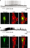
Expression of CRF1 receptor immunoreactivity (IR) on urocortin-1 (Ucn1)-responsive myenteric neurons in the guinea pig distal colon. (A) Bath application of Ucn1 (30 nM) evoked slowly activating membrane depolarization and enhanced excitability in an AH-type neuron. The neuron was traced by filling with biocytin, and later staining for biocytin and the CRF1 receptor. The Ucn1-responsive neuron with AH-type electrophysiological behaviour and Dogiel type II morphology was found to express CRF1 IR. (B) Bath application of Ucn1 (30 nM) evoked slowly activating membrane depolarization and enhanced excitability in an S-type neuron. The Ucn1-responsive S-type neuron was found to have uniaxonal morphology and express CRF1 receptor IR. Scale bars = 20 µm.
Figure 9
Figure 9
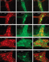
Colocalization of CRF1 receptor immunoreactivity (IR) with neurochemical codes in the myenteric plexus of the guinea pig distal colon. (A1–3) Double labelling of CRF1 receptors with anti-Hu, which labels all enteric neurons, showed that the CRF1 receptor was expressed exclusively by myenteric neurons. (B1–3) CRF1 receptor IR was colocalized with calbindin IR. (C1–3) CRF1 receptor IR was colocalized with ChAT. IR. (D1–3) CRF1 receptor IR was colocalized with nitric oxide synthase (NOS) IR. (E1–3) A small subset of CRF1 receptor-immunoreactive myenteric neurons also expressed CRF2 receptor IR. Scale bars = 20 µm.
Figure 10
Figure 10
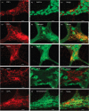
Colocalization of CRF2 receptor immunoreactivity (IR) with neurochemical markers in the myenteric plexus of the guinea pig distal colon. (A1–3) Double labelling of CRF2 receptors with anti-Hu, which labels all enteric neurons, showed expression of CRF2 receptor IR by only a small subset of neurons. (B1–3) CRF2 receptor IR was colocalized with calbindin IR. (C1–3) CRF2 receptor IR was colocalized with ChAT IR. (D1–3) CRF2 receptor IR was not colocalized with NOS IR. (E1–3) CRF2 receptor IR and synaptophysin IR were coexpressed in varicose nerve fibres. Scale bars = 20 µm.
Figure 11
Figure 11
Exposure to urocortins (Ucns) did not alter excitatory neurotransmission in the myenteric plexus of guinea-pig colon. (A) Fast excitatory postsynaptic potentials (EPSPs) evoked in an S-type myenteric neuron were unaffected by Ucn1, Ucn2 or Ucn3. (B) Slow EPSPs evoked in the same myenteric neuron were unaffected by Ucn1, Ucn2 or Ucn3. Morphology of the neuron from which the results in A and B were obtained appears in the inset. Scale bars = 20 µm.

Similar articles

Cited by

References

    1. Alexander SPH, Mathie A, Peters JA. Guide to Receptors and Channels (GRAC), 3rd edn. Br J Pharmacol. 2008;153(Suppl. 2):S1–S209. - PMC - PubMed
    1. Bale TL, Vale WW. CRF and CRF receptors: role in stress responsivity and other behaviors. Annu Rev Pharmacol Toxicol. 2004;44:525–357. - PubMed
    1. Bertrand PP, Kunze WA, Bornstein JC, Furness JB, Smith ML. Analysis of the responses of myenteric neurons in the small intestine to chemical stimulation of the mucosa. Am J Physiol. 1997;273:G422–G435. - PubMed
    1. Bertrand PP, Kunze WA, Bornstein JC, Furness JB. Electrical mapping of the projections of intrinsic primary afferent neurones to the mucosa of the guinea-pig small intestine. Neurogastroenterol Motil. 1998;10:533–541. - PubMed
    1. Bertrand PP, Kunze WA, Furness JB, Bornstein JC. The terminals of myenteric intrinsic primary afferent neurons of the guinea-pig ileum are excited by 5-hydroxytryptamine acting at 5-hydroxytryptamine-3 receptors. Neuroscience. 2000;101:459–469. - PubMed

Publication types

Substances