

Anatomical models and wax Venuses: art masterpieces or scientific craft works?
- PMID: 20002228
- PMCID: PMC2815944
- DOI: 10.1111/j.1469-7580.2009.01169.x
Anatomical models and wax Venuses: art masterpieces or scientific craft works?
Abstract
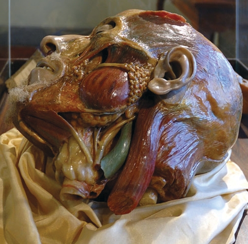
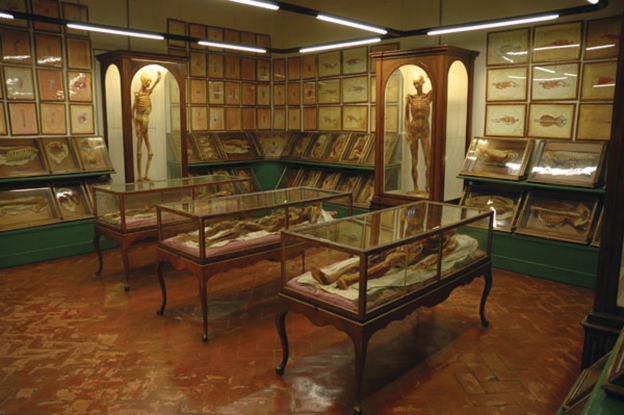
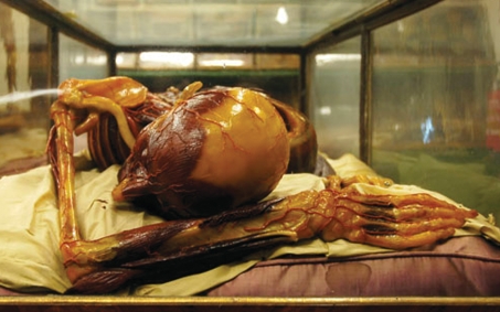
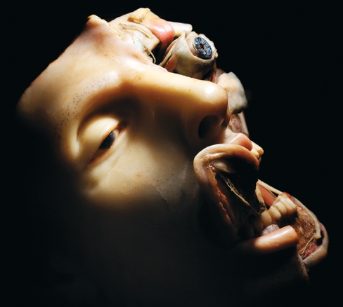
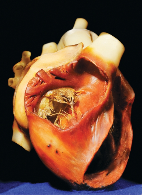
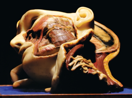
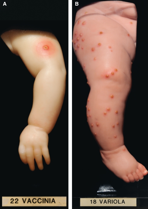
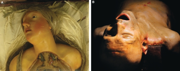
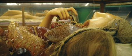
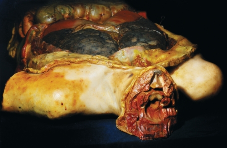
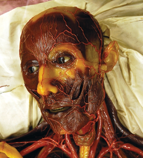
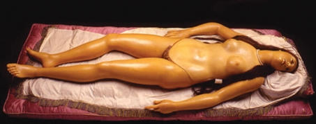
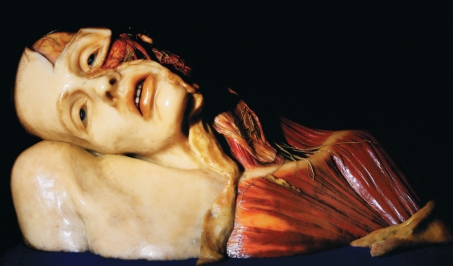
The art of wax modelling has an ancient origin but rose to prominence in 14th century Italy with the cult of votive artefacts. With the advent of Neoclassicism this art, now deemed repulsive, continued to survive in a scientific environment, where it flourished in the study of normal and pathological anatomy, obstetrics, zoology and botany. The achievement of having originated the creation of anatomical models in coloured wax must be ascribed to a joint effort undertaken by the Sicilian wax modeller Gaetano Giulio Zumbo and the French surgeon Guillaume Desnoues in the late 17th century. Interest in anatomical wax models spread throughout Europe during the 18th century, first in Bologna with Ercole Lelli, Giovanni Manzolini and Anna Morandi, and then in Florence with Felice Fontana and Clemente Susini. In England, the art of anatomical ceroplastics was brought to London from Florence by the sculptor Joseph Towne. Throughout the centuries many anatomical artists preferred this material due to the remarkable mimetic likeness obtained, far surpassing any other material. Independent of the material used, whether wood, wax or clay, anatomical models were always considered merely craft works confined to hospitals or faculties of medicine and have survived to this day only because of their scientific interest. Italian and English waxes are stylistically different but the remarkable results obtained by Susini and Towne, and the fact that some contemporary artists are again representing anatomical wax bodies in their works, makes the border that formerly separated art and craft indistinguishable.
Figures
References
-
- Antoine E. Ex voto. In: André Vauchez., editor. Encyclopedia of the Middle Ages. Cambridge: James Clarke & Co.; 2001. (e-reference edition). Distributed by Oxford University Press. Open University. 25 July 2009. Available at: http://www.oxfordreference.com.
-
- Azzaroli ML. La ceroplastica nella scienza e nell’arte. Atti del I Congresso Internazionale. Florence: Leo S. Olschki Editore; 1977. La Specola. The zoological museum of Florence University; pp. 1–22.
-
- Azzaroli Puccetti ML, Galli G, Scarani P. In: Vanitas Vanitatum, Studi sulla ceroplastica di Gaetano Giulio Zumbo. Giansiracusa P, editor. Syracuse: Lombardi; 1991.
-
- Ballestriero R. La Ceroplastica Splendore ed eclissi dal Rinascimento al Romanticismo. Academy of Fine Arts of Venice; academic year, Unpublished Thesis.
-
- Ballestriero R. Realidad y Representación en la Ceroplastíca. Faculty of Fine Arts, Complutense University Madrid; 2000. MPhil Project.
Publication types
MeSH terms
Substances
LinkOut - more resources
Full Text Sources
