. 2010 Mar;159(3):292-302.
doi: 10.1111/j.1365-2249.2009.04069.x. Epub 2009 Dec 4.

Flk-1+ mesenchymal stem cells aggravate collagen-induced arthritis by up-regulating interleukin-6

Affiliations

Flk-1+ mesenchymal stem cells aggravate collagen-induced arthritis by up-regulating interleukin-6

B Chen et al. Clin Exp Immunol. 2010 Mar.

Abstract

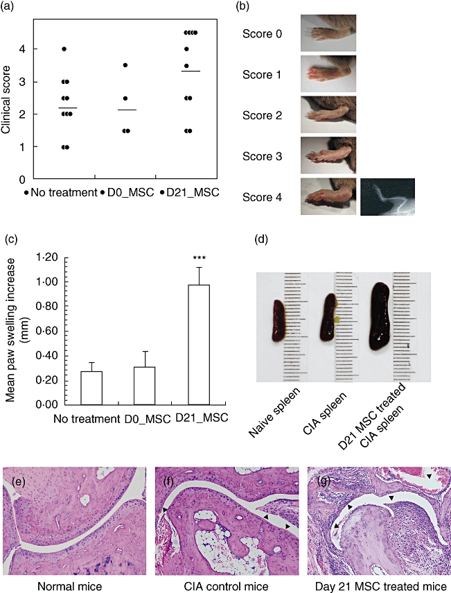
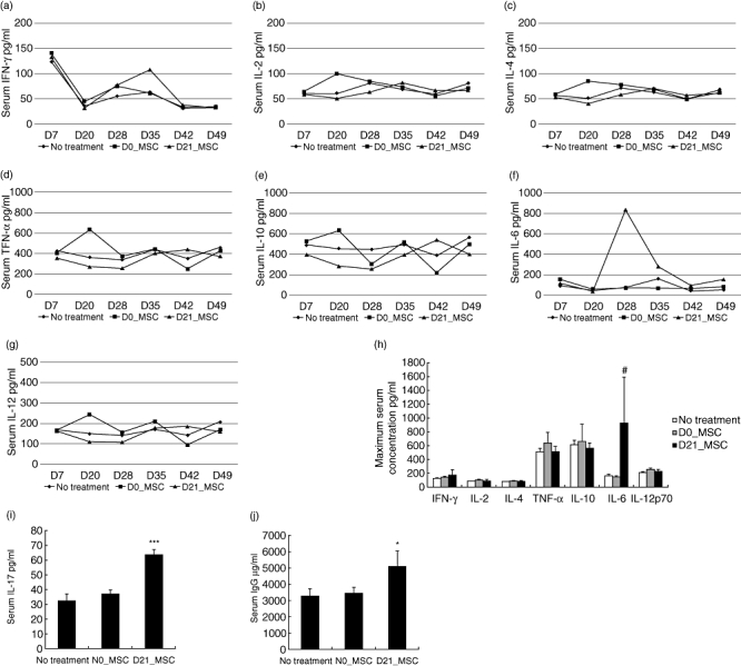
The immunomodulatory ability of mesenchymal stem cells (MSCs) may be used to develop therapies for autoimmune diseases. Flk-1(+) MSCs are a population of MSCs with defined phenotype and their safety has been evaluated in Phase 1 clinical trials. We designed this study to evaluate whether Flk-1(+) MSCs conferred a therapeutic effect on collagen-induced arthritis (CIA), an animal model of rheumatic arthritis, and to explore the underlying mechanisms. Flk-1(+) MSCs, 1-2 x 10(6), were injected into CIA mice on either day 0 or day 21. The clinical course of arthritis was monitored. Serum cytokine profile was determined by cytometric bead array kit or enzyme-linked immunosorbent assay. Flk-1(+) MSCs and splenocytes co-culture was conducted to explore the underlying mechanisms. Flk-1(+) MSCs did not confer therapeutic benefits. Clinical symptom scores and histological evaluation suggested aggravation of arthritis in mice treated with MSCs at day 21. Serum cytokine profile analysis showed marked interleukin (IL)-6 secretion immediately after MSC administration. Results of in vitro culture of splenocytes confirmed that the addition of Flk-1(+) MSCs promoted splenocyte proliferation and increased IL-6 and IL-17 secretion. Moreover, splenocyte proliferation was also enhanced in mice treated with MSCs at day 21. Accordingly, MSCs at low concentrations were found to promote lipopolysaccharide-primed splenocytes proliferation in an in vitro co-culture system. We propose that Flk-1(+) MSCs aggravate arthritis in CIA model by at least up-regulating secretion of IL-6, which favours Th17 differentiation. When Flk-1(+) MSCs are used for patients, we should be cautious about subjects with rheumatoid arthritis.


Figures

Fig. 1
Fig. 1
Immunophenotype and immunosuppressive properties of Flk-1+ mesenchymal stem cells (MSCs). Cells were cultured from murine bone marrow. (a) Immunophenotype was detected by flow cytometry. MSCs were positive for CD29, CD44, CD105 and major histocompatibility complex 1 (MHC-1) and negative for CD31, CD33, CD34, CD45, CD108, CD117 and MHC-2. (b) Cells were positive for intracellular antigen Flk-1. (c) Murine splenocytes were stimulated with either concanavalin A (ConA) or lipopolysaccharide (LPS). Cell proliferation was shown by tritiated thymidine ([3H]-TdR) incorporation and expressed in counts per minute (cpm). Addition of irradiated Flk-1+ MSCs (MSC : splenocyte = 1:10) suppressed stimulated splenocyte proliferation. (***P < 0·01).
Fig. 5
Fig. 5
Flk-1+ mesenchymal stem cells (MSCs) at low concentrations stimulate B cell proliferation in vitro. (a) Splenocytes from naive mice were cultured with irradiated Flk-1+ MSCs at different doses of Flk-1+ MSCs. Cell proliferation was measured by tritiated thymidine ([3H]-TdR) incorporation and expressed in counts per minute (cpm). (b) Concanavalin A (ConA)-stimulated splenocytes from naive mice were cultured with irradiated Flk-1+ MSCs at different doses. (c) Lipopolysaccharide (LPS)-stimulated splenocytes from naive mice were cultured with irradiated Flk-1+ MSCs at different doses. (d) Proliferation of cell from mice of the control group and the day 21 group was shown by [3H]-TdR incorporation. (e) ConA-stimulated splenocytes from collagen-induced arthritis (CIA) mice were cultured with irradiated Flk-1+ MSCs at different doses. (f) LPS-stimulated splenocytes from CIA mice were cultured with irradiated Flk-1+ MSCs at different doses (*P < 0·05 versus culture without Flk-1+ MSCs or control CIA mice; ***P < 0·01 versus culture without Flk-1+ MSCs or control CIA mice).
Fig. 2
Fig. 2
Aggravation of collagen-induced arthritis after Flk-1+ mesenchymal stem cells (MSCs) treatment at day 21. (a) Clinical scores of each group of collagen-induced arthritis (CIA) mice were evaluated according to standards described in Material and methods. (D0_MSC = day 0 group; D21_MSC = day 21 group). (b) Paws with different clinical scores. Paws of score 4 were confirmed by radiographic examination showing limb deformities and/or bone erosion. (c) Mean paw swelling increase was calculated by averaging the paw swelling increases on days 32, 35, 39, 43 and 49 (***P < 0·01 versus control group). (d) Spleens recovered from naive mice, mice in the control group and the day 21 group. (e–g) Histological sections of naive mice, CIA control mice or mice in the day 21 group were stained with haematoxylin and eosin.
Fig. 3
Fig. 3
Comparison of serum cytokine concentrations between the day 21 group, the day 0 group and control mice. (a–g) Serum samples were obtained on days 7, 20, 28, 35, 42 and 49, respectively. Serum concentrations of interleukin (IL)-2, IL-4, IL-6, IL-10, IL-12, interferon (IFN)-γ and tumour necrosis factor (TNF)-α were determined using the Murine Cytometric Bead Array Kit (BD). (h) Maximum serum cytokine concentrations during the disease progress of an individual mouse. Mice in the day 21 group showed 4·46-fold increase in serum IL-6 versus untreated collagen-induced arthritis (CIA) mice. (i–j) Serum concentrations of IL-17 and immunoglobulin (Ig)G (#P < 0·1 versus control mice; *P < 0·05 versus control mice; ***P < 0·01 versus control mice).
Fig. 4
Fig. 4
Addition of Flk-1+ mesenchymal stem cells (MSCs) to splenocyte cultures increase supernatant concentrations of interleukin (IL)-6 and IL-17. (a,c) Splenocytes from naive mice stimulated by lipopolysaccharide (LPS) were cultured with or without Flk-1+ MSCs (MSC : splenocyte = 1:10). (b,d) Splenocytes from collagen-induced arthritis (CIA) mice stimulated by LPS were cultured with or without Flk-1+ MSCs. Supernatant concentrations of IL-6 and IL-17 were determined by enzyme-linked immunosorbent assay (*P < 0·05 versus culture without Flk-1+ MSCs; ***P < 0·01 versus culture without Flk-1+ MSCs).
Fig. 6
Fig. 6
Illustration of the proposed mechanism of aggravation of arthritis by Flk-1+ mesenchymal stem cells (MSCs) treatment at day 21. Surfing interleukin (IL)-6, by enhancing T helper type 17 (Th17) and plasma cells, and the stimulatory effects of a low dose of Flk-1+ MSCs on B cell proliferation account primarily for the aggravation of collagen-induced arthritis (CIA) after Flk-1+ MSC treatment.

Similar articles

Cited by

References

    1. Fang B, Liao L, Shi M, Yang S, Zhao RC. Multipotency of Flk1CD34 progenitors derived from human fetal bone marrow. J Lab Clin Med. 2004;143:230–40. - PubMed
    1. Cao Y, Sun Z, Liao L, Meng Y, Han Q, Zhao RC. Human adipose tissue-derived stem cells differentiate into endothelial cells in vitro and improve postnatal neovascularization in vivo. Biochem Biophys Res Commun. 2005;332:370–9. - PubMed
    1. Deng W, Han Q, Liao L, et al. Engrafted bone marrow-derived flk-(1+) mesenchymal stem cells regenerate skin tissue. Tissue Eng. 2005;11:110–19. - PubMed
    1. Liu Y, Yan X, Sun Z, et al. Flk-1+ adipose-derived mesenchymal stem cells differentiate into skeletal muscle satellite cells and ameliorate muscular dystrophy in mdx mice. Stem Cells Dev. 2007;16:695–706. - PubMed
    1. Bartholomew A, Patil S, Mackay A, et al. Baboon mesenchymal stem cells can be genetically modified to secrete human erythropoietin in vivo. Hum Gene Ther. 2001;12:1527–41. - PubMed

Publication types

MeSH terms