. 2010 Apr;129(4):482-95.
doi: 10.1111/j.1365-2567.2009.03200.x. Epub 2009 Dec 2.

Activation of mannan-binding lectin-associated serine proteases leads to generation of a fibrin clot

Affiliations

Activation of mannan-binding lectin-associated serine proteases leads to generation of a fibrin clot

Krishana C Gulla et al. Immunology. 2010 Apr.

Abstract

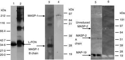
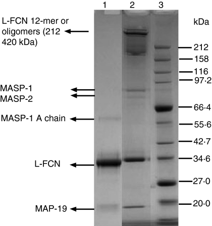
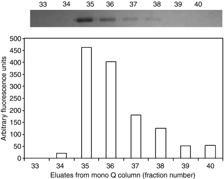
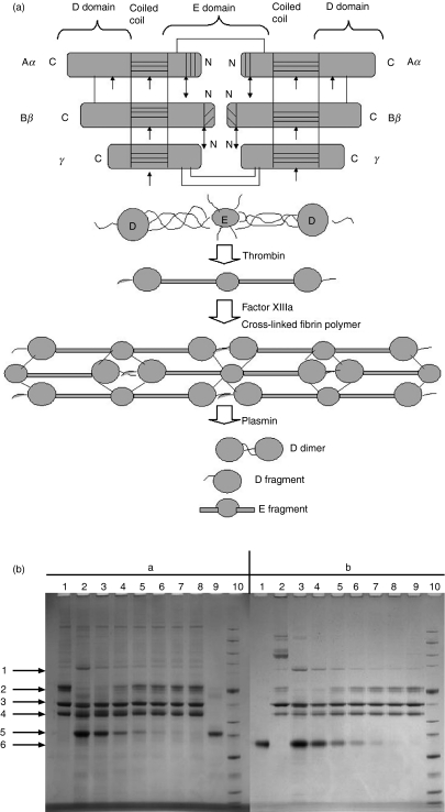
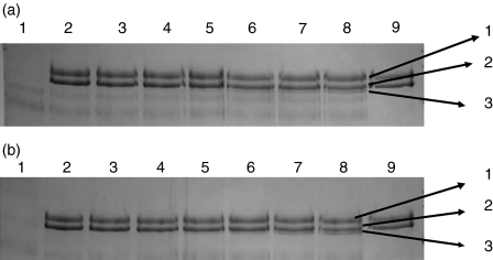
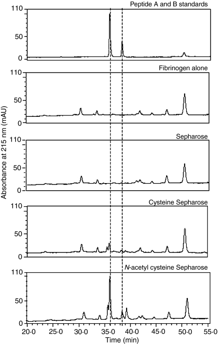
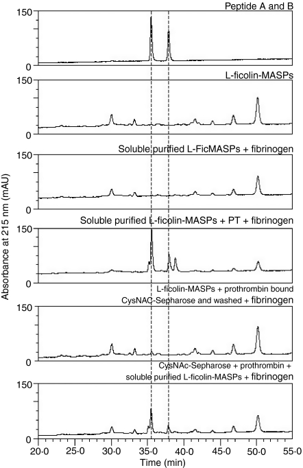
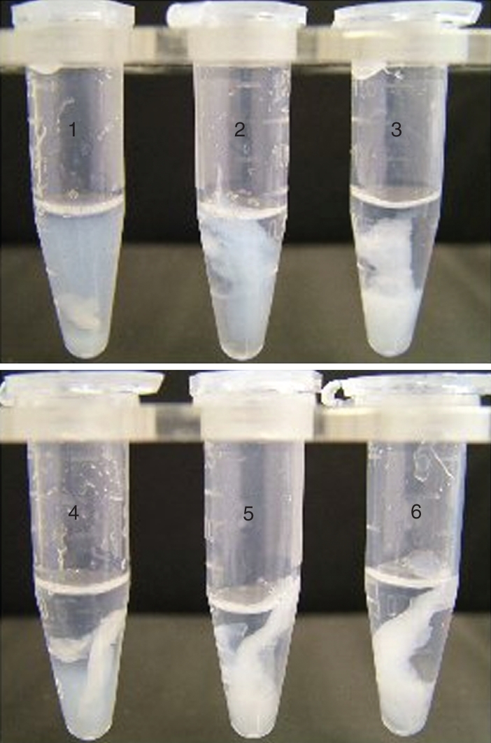
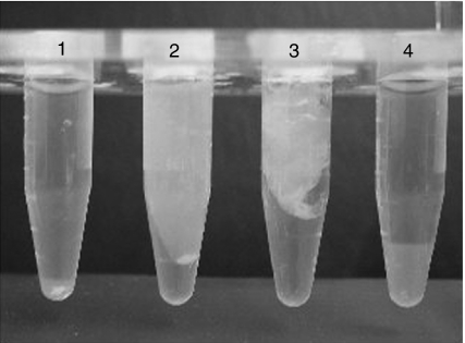
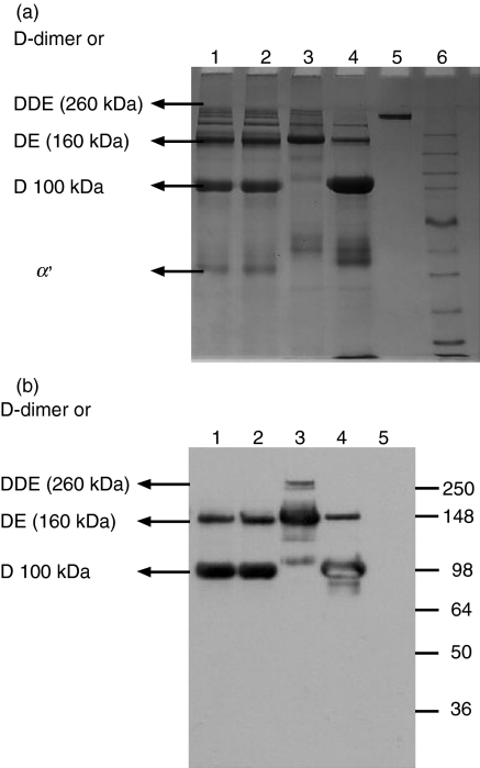
The lectin pathway of complement is activated upon binding of mannan-binding lectin (MBL) or ficolins (FCNs) to their targets. Upon recognition of targets, the MBL-and FCN-associated serine proteases (MASPs) are activated, allowing them to generate the C3 convertase C4b2a. Recent findings indicate that the MASPs also activate components of the coagulation system. We have previously shown that MASP-1 has thrombin-like activity whereby it cleaves and activates fibrinogen and factor XIII. MASP-2 has factor Xa-like activity and activates prothrombin through cleavage to form thrombin. We now report that purified L-FCN-MASPs complexes, bound from serum to N-acetylcysteine-Sepharose, or MBL-MASPs complexes, bound to mannan-agarose, generate clots when incubated with calcified plasma or purified fibrinogen and factor XIII. Plasmin digestion of the clot and analysis using anti-D-dimer antibodies revealed that the clot was made up of fibrin and was similar to that generated by thrombin in normal human plasma. Fibrinopeptides A and B (FPA and FPB, respectively) were released after fibrinogen cleavage by L-FCN-MASPs complexes captured on N-acetylcysteine-Sepharose. Studies of inhibition of fibrinopeptide release indicated that the dominant pathway for clotting catalysed by the MASPs is via MASP-2 and prothrombin activation, as hirudin, a thrombin inhibitor that does not inhibit MASP-1 and MASP-2, substantially inhibits fibrinopeptide release. In the light of their potent chemoattractant effects on neutrophil and fibroblast recruitment, the MASP-mediated release of FPA and FPB may play a role in early immune activation. Additionally, MASP-catalysed deposition and polymerization of fibrin on the surface of micro-organisms may be protective by limiting the dissemination of infection.


Figures

Figure 2
Figure 2
Western blots of the L-FCN preparation. Western blots were probed with anti-L-FCN (lanes 1 and 2), anti-MASP-1 (lanes 3 and 4) and anti-MASP-2 (lanes 5 and 6) under reducing and non-reducing conditions, respectively. As can be observed, L-FCN ran as a single polypeptide band of 35 kDa under reducing conditions while under non-reducing conditions it formed a ladder of bands. This running pattern is characteristic of a homo-oligomeric protein such as FCN. When a similar blot was probed with anti-MASP-1 (B-chain specific) antibodies only the MASP-1 B chain was detectable under reducing conditions, while the A and B chain complex could be detected under non-reducing conditions. On the blot probed with anti-MASP-2 (A-chain specific) antibodies both the MASP-2 A chain and MAp19 could be detected under reducing conditions while only the A and B chain complex could be detected in the non-reducing conditions.
Figure 1
Figure 1
Sodium dodecyl sulphate–polyacrylamide gel electrophoresis (SDS-PAGE) pattern of L-FCN-MASPs. Purified L-FCN-MASPs were separated under reducing (lane 1) and non-reducing (lane 2) conditions. Markers: lane 3. Gels were stained with Coomassie brilliant blue.
Figure 3
Figure 3
Cleavage of a synthetic peptide FGR-AMC by L-FCN-MASPs in mono Q eluates. The top panel shows the amount of 35-kDa L-FCN present in fractions eluted from a mono Q column, as indicated by sodium dodecyl sulphate–polyacrylamide gel electrophoresis (SDS-PAGE). The chart shows the corresponding proteolytic activity of the L-FCN-associated MASP, following capture of the L-ficolin on an N-acetyl bovine serum albumin (BSA) surface. MASP activity in the individual fractions was quantified by measuring the fluorescence increase after cleavage of the peptide derivative FGR-AMC (Presanis et al., 2004). The results indicate that the L-FCN detected by SDS-PAGE was indeed complexed with active MASPs.
Figure 4
Figure 4
(a) Fibrin polymerization and lysis. The top panel shows polypeptide organization of fibrinogen. The area with horizontal lines is the coiled coil region. The area with vertical lines is fibrinopeptide A. The area with diagonal lines is fibrinopeptide B. Double arrows indicate the thrombin cleavage site. Single arrows indicate plasmin cleavage sites. The middle panel shows the domain organization of fibrinogen. The bottom panel shows cross-linking of the fibrin monomer. The E domain binds to the holes on up to four D domains, forming a long fibrous latticework. The clot is then stabilized through cross-linking. The clot can be degraded, yielding different degradation products if it has been cross-linked. The D fragment is released when the clot is not cross-linked by factor XIIIIa. (b) Fibrinogen cleavage by purified lectin-MASPs. Fibrinogen was incubated with various concentrations of purified lectin-MASPs and subjected to reducing sodium dodecyl sulphate–polyacrylamide gel electrophoresis (SDS-PAGE). Arrows indicate (1) the γ-dimer, (2) the α-chain, (3) the β-chain, (4) the γ-chain, (5) L-FCN and (6) mannan-binding lectin (MBL). Panels (a) and (b) show the results obtained with L-FCN-MASPs and MBL-MASPs, respectively. (a) Lane 1, fibrinogen (5 μg) incubated without L-FCN-MASPs at 37° for 14 hr; lanes 2–8, with 3·5, 1·5, 0·75, 0·4, 0·2, 0·1,0 05 and 0·025 μg of L-FCN-MASPs; lane 9, with 1·25 μg of L-FCN-MASPs; lane 10, marker. (b) Lane 1, MBL-MASPs (1 ug); lane 2, fibrinogen (1 ug); lanes 3–9, fibrinogen (1 ug) incubated with 3·0, 1·5, 0·75, 0·375, 0·18, 0·09 and 0·046 ug of MBL-MASPs; lane 10, marker. The results show that both L-FCN and MBL exhibit a dose-dependent cross-linking of the fibrinogen γ-chains, resulting in the generation of the γ-dimer as well as degradation of the α-chain. Additionally, a small change in the size of the β-chain could be observed, indicating the release of fibrinopeptide B.
Figure 5
Figure 5
Factor XIII cleavage by purified lectin mannan-binding lectin-associated serine proteases (MASPs). Factor XIII was incubated with L-FCN-MASPs and mannan-binding lectin (MBL) MASPs for different times at 37° and subjected to reducing sodium dodecyl sulphate–polyacrylamide gel electrophoresis (SDS-PAGE). Panels (a) and (b) show the results obtained with L-FCN-MASPs and MBL-MASPs, respectively. Arrows indicate (1) the B chain, (2) the A chain, (3) the cleaved A chain. (a) Lane 1, L-FCN-MASPs; lanes 2–8, factor XIII + L-FCN-MASPs incubated for 0 min, 30 min, 1 hr, 2 hr, 4 hr, 6 hr and 8 hr, respectively; lane 9, factor XIII alone incubated for 8 hr. (b) Factor XIII (1 μg) incubated with 2·5 μg of purified MBL-MASPs for different time intervals at 37° and subjected to SDS-PAGE under reducing conditions. Lane 1, MBL-MASPs; lanes 2–8, factor XIII + MBL-MASPs incubated for 0 min, 30 min, 1 hr, 2 hr, 4 hr, 6 hr and 8 hr; lane 9, factor XIII incubated for 8 hr. Arrows indicate (1) the B chain, (2) the A chain and (3) the cleaved A chain.
Figure 6
Figure 6
Fibrinopeptide release by ficolin MASPs trapped on various matrices. Fibrinopeptides were released following cleavage of fibrinogen by L-FCN-MASPs as described in the Materials and methods.
Figure 7
Figure 7
Effect of prothrombin addition to purified L-FCN-MASPs in fibrinopeptide release. Fibrinopeptides were released following cleavage of fibrinogen by L-FCN-MASP in the presence and absence of prothrombin, as described in the Materials and methods.
Figure 8
Figure 8
Generation of a clot from fibrinogen in the presence or absence of factor XIII by MASPs captured on CysNAc-Sepharose or mannan-binding lectin (MBL) MASPs captured on mannan-agarose. 1, CysNAc-Sepharose + fibrinogen + iodoacetamide; 2, CysNAc-Sepharose + fibrinogen + factor XIII; 3, CysNAc-Sepharose + soybean trypsin inhibitor (SBTI) + fibrinogen + factor XIII; 4, mannan-agarose + fibrinogen + iodoacetamide; 5, mannan-agarose + fibrinogen + factor XIII; 6, mannan-agarose + SBTI + fibrinogen + factor XIII.
Figure 9
Figure 9
Clot generation by mannan-binding lectin (MBL)/MASPs bound to their respective matrices after incubation with diluted plasma. Lane 1, underivatized Sepharose; lane 2, CysNAc-Sepharose; lane 3, mannan-agarose; lane 4, mannan-agarose incubated with diluted plasma in the presence of 20 mm ethylenediaminetetraacetic acid (EDTA).
Figure 10
Figure 10
(a) Comparison of plasmin digests of generated clots. Lane 1, plasmin digest of a clot generated from plasma by CysNAc-Sepharose; lane 2, plasmin digest of a clot generated from plasma by mannan-Sepharose; lane 3, plasmin digest of a clot generated by thrombin and fibrinogen in the presence of factor XIII; 4, plasmin digest of a clot generated by thrombin and fibrinogen in the absence of factor XIII; 5, fibrinogen; 6, marker. (b) Western blots of plasmin digests of the generated clots. Lane 1, plasmin digest of a clot generated from plasma by CysNAc-Sepharose; lane 2, plasmin digest of a clot generated from plasma by mannan-Sepharose; lane 3, plasmin digest of a clot generated by thrombin and fibrinogen in the presence of factor XIII; lane 4, plasmin digest of a clot generated by thrombin and fibrinogen in the absence of factor XIII; lane 5, fibrinogen; lane 6, marker.

Similar articles

Cited by

References

    1. Fujita T. Evolution of the lectin-complement pathway and its role in innate immunity. Nat Rev Immunol. 2002;2:346–53. - PubMed
    1. Gal P, Dobo J, Zavodszky P, Sim RB. Early complement proteases: C1r, C1s and MASPs. A structural insight into activation and functions. Mol Immunol. 46:2745–52. - PubMed
    1. Mayilyan KR, Presanis JS, Arnold JN, Hajela K, Sim RB. Heterogeneity of MBL- MASP complexes. Mol Immunol. 2006;43:1286–92. - PubMed
    1. Mayilyan KR, Presanis JS, Arnold JN, Sim RB. Discrete MBL-MASP complexes show wide inter-individual variability in concentration: data from UK vs Armenian populations. Int J Immunopathol Pharmacol. 2006;19:567–80. - PubMed
    1. Bhakdi S, Tranum-Jensen J. Complement lysis: a hole is a hole. Immunol Today. 1991;12:318–21. - PubMed

Publication types

MeSH terms

LinkOut - more resources