

Calcium-binding proteins in skeletal muscles of the mdx mice: potential role in the pathogenesis of Duchenne muscular dystrophy
- PMID: 20002835
- PMCID: PMC2812729
- DOI: 10.1111/j.1365-2613.2009.00688.x
Calcium-binding proteins in skeletal muscles of the mdx mice: potential role in the pathogenesis of Duchenne muscular dystrophy
Abstract
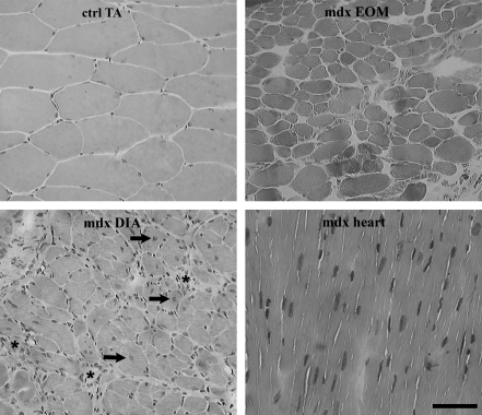
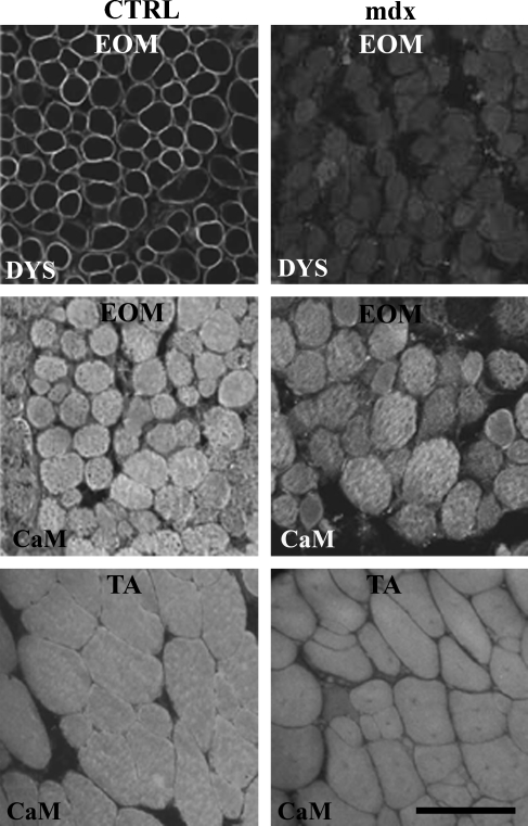
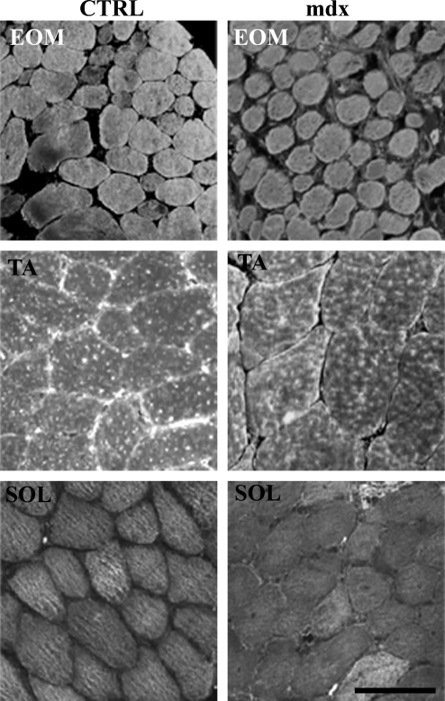
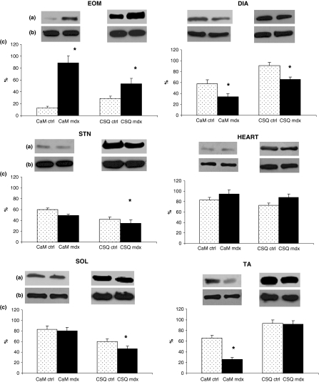
Duchenne muscular dystrophy is one of the most common hereditary diseases. Abnormal ion handling renders dystrophic muscle fibers more susceptible to necrosis and a rise in intracellular calcium is an important initiating event in dystrophic muscle pathogenesis. In the mdx mice, muscles are affected with different intensities and some muscles are spared. We investigated the levels of the calcium-binding proteins calsequestrin and calmodulin in the non-spared axial (sternomastoid and diaphragm), limb (tibialis anterior and soleus), cardiac and in the spared extraocular muscles (EOM) of control and mdx mice. Immunoblotting analysis showed a significant increase of the proteins in the spared mdx EOM and a significant decrease in the most affected diaphragm. Both proteins were comparable to the cardiac muscle controls. In limb and sternomastoid muscles, calmodulin and calsequestrin were affected differently. These results suggest that differential levels of the calcium-handling proteins may be involved in the pathogenesis of myonecrosis in mdx muscles. Understanding the signaling mechanisms involving Ca(2+)-calmodulin activation and calsequestrin expression may be a valuable way to develop new therapeutic approaches to the dystrophinopaties.
Figures
Similar articles
-
Isobaric Tagging-Based Quantification for Proteomic Analysis: A Comparative Study of Spared and Affected Muscles from mdx Mice at the Early Phase of Dystrophy.PLoS One. 2013 Jun 18;8(6):e65831. doi: 10.1371/journal.pone.0065831. Print 2013. PLoS One. 2013. PMID: 23823696 Free PMC article.
-
Changes in calsequestrin, TNF-α, TGF-β and MyoD levels during the progression of skeletal muscle dystrophy in mdx mice: a comparative analysis of the quadriceps, diaphragm and intrinsic laryngeal muscles.Int J Exp Pathol. 2015 Oct;96(5):285-93. doi: 10.1111/iep.12142. Epub 2015 Oct 30. Int J Exp Pathol. 2015. PMID: 26515458 Free PMC article.
-
Stretch-activated calcium channel protein TRPC1 is correlated with the different degrees of the dystrophic phenotype in mdx mice.Am J Physiol Cell Physiol. 2011 Dec;301(6):C1344-50. doi: 10.1152/ajpcell.00056.2011. Epub 2011 Sep 7. Am J Physiol Cell Physiol. 2011. PMID: 21900691
-
Pharmacological control of cellular calcium handling in dystrophic skeletal muscle.Neuromuscul Disord. 2002 Oct;12 Suppl 1:S155-61. doi: 10.1016/s0960-8966(02)00095-0. Neuromuscul Disord. 2002. PMID: 12206810 Review.
-
Proteomic profiling of x-linked muscular dystrophy.J Muscle Res Cell Motil. 2009 Dec;30(7-8):267-9. doi: 10.1007/s10974-009-9197-6. J Muscle Res Cell Motil. 2009. PMID: 20082121 Review.
Cited by
-
The Effect of Vitamin D Supplementation on Skeletal Muscle in the mdx Mouse Model of Duchenne Muscular Dystrophy.Sports (Basel). 2019 Apr 26;7(5):96. doi: 10.3390/sports7050096. Sports (Basel). 2019. PMID: 31035483 Free PMC article.
-
Abnormal Calcium Handling in Duchenne Muscular Dystrophy: Mechanisms and Potential Therapies.Front Physiol. 2021 Apr 9;12:647010. doi: 10.3389/fphys.2021.647010. eCollection 2021. Front Physiol. 2021. PMID: 33897454 Free PMC article. Review.
-
Isobaric Tagging-Based Quantification for Proteomic Analysis: A Comparative Study of Spared and Affected Muscles from mdx Mice at the Early Phase of Dystrophy.PLoS One. 2013 Jun 18;8(6):e65831. doi: 10.1371/journal.pone.0065831. Print 2013. PLoS One. 2013. PMID: 23823696 Free PMC article.
-
Calsequestrin 2 and arrhythmias.Am J Physiol Heart Circ Physiol. 2012 Mar 15;302(6):H1250-60. doi: 10.1152/ajpheart.00779.2011. Epub 2011 Dec 23. Am J Physiol Heart Circ Physiol. 2012. PMID: 22198169 Free PMC article. Review.
-
Multiple LEDT wavelengths modulate the Akt signaling pathways and attenuate pathological events in mdx dystrophic muscle cells.Photochem Photobiol Sci. 2022 Jul;21(7):1257-1272. doi: 10.1007/s43630-022-00216-0. Epub 2022 Apr 5. Photochem Photobiol Sci. 2022. PMID: 35380391
References
-
- Alderton JM, Steinhardt RA. Calcium influx through calcium leak channels is responsible for the elevated levels of calcium-dependent proteolysis in dystrophic myotubes. J. Biol. Chem. 2000;275:9452–9460. - PubMed
-
- Anderson JT, Rogers RP, Jarrett HW. Ca2+-calmodulin binds to the carboxyl-terminal domain of dystrophin. J. Biol. Chem. 1996;271:6605–6610. - PubMed
-
- Andrade FH, Porter JD, Kaminski HJ. Eye muscle sparing by the muscular dystrophies: lessons to be learned? Micros. Res. Tec. 2000;48:192–203. - PubMed
-
- Angus LM, Chakkalakal JV, Mejat A, et al. Calcineurin/NFAT signaling, together with GABP and PGC-1alpha, drives utrophin gene expression at the neuromuscular junction. Am. J. Physiol. Cell Physiol. 2005;289:C908–C917. - PubMed
-
- Beard NA, Laver DR, Dulhunty AF. Calsequestrin and the calcium release channel of skeletal and cardiac muscle. Prog. Biophys. Mol. Biol. 2004;85:33–69. - PubMed
Publication types
MeSH terms
Substances
LinkOut - more resources
Full Text Sources
Miscellaneous
