. 2009 Dec 9:7:146.
doi: 10.1186/1477-7827-7-146.

Transforming growth factor-beta inhibits aromatase gene transcription in human trophoblast cells via the Smad2 signaling pathway

Affiliations

Transforming growth factor-beta inhibits aromatase gene transcription in human trophoblast cells via the Smad2 signaling pathway

Hong Zhou et al. Reprod Biol Endocrinol. .

Abstract

Background: Transforming growth factor-beta (TGF-beta) is known to exert multiple regulatory functions in the human placenta, including inhibition of estrodial production. We have previously reported that TGF-beta1 decreased aromatase mRNA levels in human trophoblast cells. The objective of this study was to investigate the molecular mechanisms underlying the regulatory effect of TGF-beta1 on aromatase expression.

Methods: To determine if TGF-beta regulates aromatase gene transcription, several reporter constructs containing different lengths of the placental specific promoter of the human aromatase gene were generated. JEG-3 cells were transiently transfected with a promoter construct and treated with or without TGF-beta1. The promoter activity was measured by luciferase assays. To examine the downstream signaling molecule mediating the effect of TGF-beta on aromatase transcription, cells were transiently transfected with dominant negative mutants of TGF-beta type II (TbetaRII) and type I receptor (ALK5) receptors before TGF-beta treatment. Smad2 activation was assessed by measuring phophorylated Smad2 protein levels in cytosolic and nuclear fractions. Smad2 expression was silenced using a siRNA expression construct. Finally, aromatase mRNA half-life was determined by treating cells with actinomycin D together with TGF-beta1 and measuring aromatase mRNA levels at various time points after treatment.

Results and discussion: TGF-beta1 inhibited the aromatase promoter activity in a time- and dose-dependent manner. Deletion analysis suggests that the TGF-beta1 response element resides between -422 and -117 nucleotides upstream from the transcription start site where a Smad binding element was found. The inhibitory effect of TGF-beta1 was blocked by dominant negative mutants of TbetaRII and ALK5. TGF-beta1 treatment induced Smad2 phosphorylation and translocation into the nucleus. On the other hand, knockdown of Smad2 expression reversed the inhibitory effect of TGF-beta1 on aroamtase transcription. Furthermore, TGF-beta1 accelerated the degradation of aromatase mRNA.

Conclusion: Our results demonstrate that TGF-beta1 exerts regulatory effects on aromatase gene at both transcriptional and post-transcriptional levels. The transcriptional regulation of aromatase gene by TGF-beta1 is mediated by the canonical TGF-beta pathway involving TbetaRII, ALK5 and Smad2. These findings further support the role of TGF-beta1 in regulating human placental functions and pregnancy.


Figures
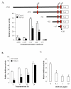
Figure 1
Figure 1
Placental aromatase promoter constructs and the effect of TGF-β1 on promoter activities. A) Various lengths (-2538 to -117) of the 5' flanking region of the placental specific exon 1 were cloned into a luciferase reporter construct pGL3 basic. Smad binding elements (SBE) are shown at the red box region. JEG-3 cells cultured in 6-well plates were transfected with these luciferase constructs (1 μg) and then treated with or without TGF-β1 (1 ng/ml) for 6 h. B) Cells were transfected with Arom -714 construct and then treated with TGF-β1 (1 ng/ml) for the duration as indicated. C) Cells were transfected with Arom -714 - and then treated with different concentrations of TGF-β1 for 6 h. In these experiments, a β-galactosidase expression vector (0.5 μg) was co-transfected into cells for normalizing transfection efficiency. Relative luciferase activity was calculated as the ratio of luciferase/β-gal. Data are mean ± SEM of three replicates from one experiment. The experiment has been repeated twice with similar results. *, P < 0.05 verse empty vector control.
Figure 2
Figure 2
TGF-β1 acts through TβRII and ALK5 to inhibit aromatase transcription. A) Cells were transfected with Arom -714 construct, the control vector pCMV5 or dominant negative TβRII. At 24 h after transfection, they were treated with the control medium or TGF-β1 (1 ng/ml). B) Cells were transfected with Arom -714 construct, control vector pCMV5, the dominant negative ALK5 (ALK5-kd) and the constitutively active ALK5 (ALK5-ca). A β-galactosidase expression vector (0.5 μg) was simultaneously co-transfected into cells for normalizing transfection efficiency.
Figure 3
Figure 3
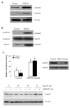
Smad2 is involved in TGF-β1-inhibited aromatase transcription. A) TGF-β1 induced Smad2 phosphorylation. Cells were treated with TGF-β1 (1 ng/ml) for 1 h and Smad2 and Smad3 phosphorylation was determined by Western blot analysis using specific antibodies. β-actin was used for the loading control. B) TGF-β1 induced Smad2 nuclear translocation. Cells were treated with TGF-β1 as above and cytoplamic and nuclear proteins were analyzed for the phosphorylated Smad2 levels. C) Validation of Smad2 siRNA. Cells were transfected with 2 μg of pSuper empty vector or Smad2 siRNA (siSmad2) cloned in pSuper. At various time points after transfection, cell lysates were prepared and probed for Smad2. D) Smad2 siRNA reversed the TGF-β1-inhibited aromatase transcription. Cells were transfected with pSuper or pSuper carrying Smad2 siRNA. Smad2 siRNA blocked the effect of TGF-β1 on aromatase transcription. Knockdown of Smad2 by its siRNA was confirmed by Western blotting.
Figure 4
Figure 4
TGF-β1 decreased aromatase mRNA stability. Cells seated in 24-well plates were treated with 2 μg/ml actinomycin D alone or in combination with TGF-β1 (1 ng/ml). Cells were harvested at time 0 (pretreatment), 6, 12, 24, 36 and 48 h after treatment with TGF-β1. Aromatase mRNA levels were determined by semi-quantitative PCR. A) Representative results from one experiment. B) The half-life (T1/2) of aromatase transcript is defined as the time required for aromatse mRNA levels to drop to 50% of its starting values (0 time point). In this study, the T1/2 value of aromatse mRNA was reduced from 36 h in the control to 20 h in the treatment groups with TGF-β1. Data are mean ± SEM of three experiments. *, P < 0.05 verse the time matched control.
Figure 5
Figure 5
Proposed model of TGF-β action in estrogen production in trophoblast cells. TGF-β binds to its serine/threonine receptor complex (TβRII and ALK5) to activate Smad2. The phosphorylated Smad2 forms a complex with Smad4 and translocate to the nucleus, where the complex could interact with other transcription factors (TF) and co-repressors to inhibit aromatase gene transcription. TGF-β also decreases aromatase mRNA stability, which contributes to a decrease in aromatase mRNA levels. The decrease in aromatase mRNA may lead to a decrease in aromatase activity and thus estrogen biosynthesis in trophoblast cells.

Similar articles

Cited by

References

    1. Knight PG, Glister C. TGF-beta superfamily members and ovarian follicle development. Reproduction. 2006;132(2):191–206. doi: 10.1530/rep.1.01074. - DOI - PubMed
    1. Peng C. The TGF-beta superfamily and its roles in the human ovary and placenta. J Obstet Gynaecol Can. 2003;25(10):834–844. - PubMed
    1. Jones RL, Stoikos C, Findlay JK, Salamonsen LA. TGF-beta superfamily expression and actions in the endometrium and placenta. Reproduction. 2006;132(2):217–232. doi: 10.1530/rep.1.01076. - DOI - PubMed
    1. Schaffer L, Scheid A, Spielmann P, Breymann C, Zimmermann R, Meuli M, Gassmann M, Marti HH, Wenger RH. Oxygen-regulated expression of TGF-beta 3, a growth factor involved in trophoblast differentiation. Placenta. 2003;24(10):941–950. doi: 10.1016/S0143-4004(03)00166-8. - DOI - PubMed
    1. Luo S, Yu H, Wu D, Peng C. Transforming growth factor-beta1 inhibits steroidogenesis in human trophoblast cells. Mol Hum Reprod. 2002;8(4):318–325. doi: 10.1093/molehr/8.4.318. - DOI - PubMed

Publication types

MeSH terms

LinkOut - more resources