. 2009 Dec 10:2:243.
doi: 10.1186/1756-0500-2-243.

Crude subcellular fractionation of cultured mammalian cell lines

Affiliations

Crude subcellular fractionation of cultured mammalian cell lines

Paul Holden et al. BMC Res Notes. .

Abstract

Background: The expression and study of recombinant proteins in mammalian culture systems can be complicated during the cell lysis procedure by contaminating proteins from cellular compartments distinct from those within which the protein of interest resides and also by solubility issues that may arise from the use of a single lysis buffer. Partial subcellular fractionation using buffers of increasing stringency, rather than whole cell lysis is one way in which to avoid or reduce this contamination and ensure complete recovery of the target protein. Currently published protocols involve time consuming centrifugation steps which may require expensive equipment and commercially available kits can be prohibitively expensive when handling large or multiple samples.

Findings: We have established a protocol to sequentially extract proteins from cultured mammalian cells in fractions enriched for cytosolic, membrane bound organellar, nuclear and insoluble proteins. All of the buffers used can be made inexpensively and easily and the protocol requires no costly equipment. While the method was optimized for a specific cell type, we demonstrate that the protocol can be applied to a variety of commonly used cell lines and anticipate that it can be applied to any cell line via simple optimization of the primary extraction step.

Conclusion: We describe a protocol for the crude subcellular fractionation of cultured mammalian cells that is both straightforward and cost effective and may facilitate the more accurate study of recombinant proteins and the generation of purer preparations of said proteins from cell extracts.


Figures

Figure 1
Figure 1
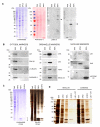
Optimization of digitonin concentration for the extraction of cytosolic proteins. (A) HEK293 cells were plated at a density of 4 × 10⁵ cells per well of a 12 well plate and were harvested 48 hours later. Cells were then lysed in 400 μl of buffer containing digitonin at the concentration indicated. Following centrifugation and collection of the supernatant the remaining cell pellet was further extracted in NP40 lysis buffer. An aliquot of each extract was then analysed by 4-12% SDS PAGE (Invitrogen #NP0322) followed by staining with Coomassie blue. (B) The concentration of protein in each extract was determined by colorimetric protein assay (Pierce #23232) and the results were plotted to demonstrate that total protein extracted was the same regardless of the starting concentration of digitonin. (C) Extracted proteins from each fraction were analyzed by Western blot using an anti GAPDH antibody (NOVUS Biologicals #300-221B) as a marker of the cytosol and an anti BiP antibody (SIGMA # G918) as a marker of the endoplasmic reticulum.
Figure 2
Figure 2
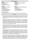
Crude subcellular fractionation protocol. Recipes to prepare the required buffers are given at the top of the figure along with additional requirements for the protocol. The protocol is then listed step by step below.
Figure 3
Figure 3
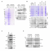
Solubilization and extraction of nuclei and insoluble proteins. (A) HEK293 cells were plated at a density of 4 × 10⁵ cells per well of a 12 well plate and processed according to the protocol in Figure 2. An aliquot of each extract was then analyzed by 4-12% SDS PAGE followed by staining with Coomassie blue or by Western blotting and probing with antibodies to the nuclear membrane protein Lamin A (SIGMA #L1293) or the ER membrane protein TRAM both highlighted by arrowheads. (B) Extracts generated in (A) were analyzed by 8 or 12% SDS PAGE followed by Western blotting and probing with antibodies to various cytosolic; Akt, Cell Signaling Technology (CST #9272), GSK-3β, (CST #9315), Caspase-3 (CST #9662) and γ-Tubulin (Santa Cruz Biotechnology, sc-7396), Organellar; Calnexin (SIGMA #C4731), Calreticulin (Stressgen #SPA-061), VDAC/Porin (SIGMA #V2139) and nuclear; p53 (CST #9282), RAS (Upstate Biotechnology #05-516) and Histone H3 (ABCAM #ab1791) proteins as indicated or with anti V5 antibody (Invitrogen #R96025) in the case of ERGIC-53. (C) Equal volumes of extracts were analyzed by 4-12% SDS-PAGE followed by staining with Coomassie blue and then silver staining. The bracket alongside the Coomassie stained gel shows the position of Histones in the RIPA nuclear extract and the asterisk next to the silver stained gel denotes a protein band unique to the E-RIPA extract. (D) The subcellular fractionation protocol was carried out in full and in duplicate. In one experiment the RIPA and E-RIPA extracts were prepared as a single extract as described in step 5 of the protocol (Combined). In both cases following the E-RIPA extract any remaining pellet was extracted by boiling in LSB + DTT.
Figure 4
Figure 4
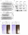
Demonstration of the advantages of crude fractionation over whole cell lysis and scale up of the approach. (A) HEK293 cells were plated at a density of 4 × 10⁵ cells per well of a 12 well plate and subsequently lysed directly in NP40 or RIPA lysis buffer or processed according to the protocol in Figure 2 (Sequential). An aliquot of each extract was then analyzed by 4-12% SDS PAGE followed by staining with Coomassie blue or by Western blotting and probing with antibodies to various marker proteins of specific intracellular organelles as previously. (B) HEK293 cells were plated at a density of 4 × 10⁵ cells per well of a 12 well dish and then transfected the next day in triplicate with 400 ng of a construct encoding V5/6×-HIS tagged ERGIC-53. Cells were extracted 48 hours later either directly in RIPA buffer, directly in NP40 buffer or according to our extraction protocol. Proteins were then partially purified from each extract using Nickel resin (SIGMA #P6611) and eluted proteins were analyzed by 10% SDS PAGE followed by staining with Coomassie blue or by Western blotting and probing with the anti V5 antibody. (C) HEK293 cells were plated at a density of 4 × 10⁵ cells per well of a 12 well dish and then transfected the next day in with 1600 ng of pcDNA6/V5-His (Invitrogen) or constructs encoding WT or MUT COMP. Cells were harvested 48 hours later and extracted according to our protocol. (D) HEK293 cells were plated at a density of 4 × 10⁵ cells per well of a 12 well dish or 8.4 × 10⁵ cells per well of a 35 mm dish and processed according to the protocol in Figure 2. An aliquot of each extract was then analyzed by 4-12% SDS PAGE followed by Western blotting and probing with antibodies to GAPDH, BiP and Lamin A.
Figure 5
Figure 5
Application of the approach to other cell lines. (A) HT1080 cells (2 × 10⁵) and HeLa cells (1 × 10⁵) were plated in the wells of a 12 well plate and the digitonin concentration required for optimal extraction of the cytosol was determined as in Figure1. (B) The remainder of the protocol from Figure 2 was then applied using 100 μg/ml digitonin as the optimal cytosolic extract in each case. An aliquot of each extract was then analyzed by 4-12% SDS PAGE followed by Western blotting and probing with antibodies to GAPDH, BiP and Histone H3 (Used as an alternative nuclear marker due to weakness of the Lamin A signal in HeLa cells). (C) Samples were analyzed by 4-12% SDS PAGE followed by staining with Coomassie blue and then silver staining showing the difference in protein composition of each sample. The bracket alongside the gels marks the position of Histones in the RIPA nuclear extract and the asterisk next to the silver stained gels denotes protein bands unique to the E-RIPA extracts.

Similar articles

Cited by

References

    1. Mooney RA. Use of digitonin-permeabilized adipocytes for cAMP studies. Methods in Enzymology. 1988;159:193–202. full_text. - PubMed
    1. Schulz I. Permeabilizing cells: some methods and applications for the study of intracellular processes. Methods in Enzymology. 1990;192:280–300. full_text. - PubMed
    1. Hide M, Ritleng A, Brizard J, Monte-Allegre A, Sereno D. Leishmania infantum: tuning digitonin fractionation for comparative proteomic of the mitochondrial protein content. Parasitology Research. 2008;103:989–992. doi: 10.1007/s00436-008-1062-9. - DOI - PubMed
    1. Foucher AL, Papadopoulou B, Ouellette M. Prefractionation by digitonin extraction increases representation of the cytosolic and intracellular proteome of Leishmania infantum. Journal of Proteome Research. 2006;5:1741–50. doi: 10.1021/pr060081j. - DOI - PubMed
    1. Huang Z, Li G, Pei W, Sosa LA, Niu L. Enhancing protein expression in single HEK 293 cells. Journal of Neuroscience Methods. 2005;142:159–166. doi: 10.1016/j.jneumeth.2004.09.009. - DOI - PubMed

LinkOut - more resources