Overexpression of inhibitor of DNA-binding (ID)-1 protein related to angiogenesis in tumor advancement of ovarian cancers
- PMID: 20003244
- PMCID: PMC2796680
- DOI: 10.1186/1471-2407-9-430
Overexpression of inhibitor of DNA-binding (ID)-1 protein related to angiogenesis in tumor advancement of ovarian cancers
Abstract
Background: The inhibitor of DNA-binding (ID) has been involved in cell cycle regulation, apoptosis and angiogenesis. This prompted us to study ID functions in tumor advancement of ovarian cancers.
Methods: Sixty patients underwent surgery for ovarian cancers. In ovarian cancers, the levels of ID-1, ID-2 and ID-3 mRNAs were determined by real-time reverse transcription-polymerase chain reaction. The histoscore with the localization of ID-1 was determined by immunohistochemistry. Patient prognosis was analyzed with a 36-month survival rate. Microvessel counts were determined by immunohistochemistry for CD34 and factor VIII-related antigen.
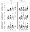
Results: ID-1 histoscores and mRNA levels both significantly (p < 0.001) increased in ovarian cancers according to clinical stage, regardless of histopathological type. Furthermore, 30 patients with high ID-1 expression had a lower survival rate (53%) compared to patients with low ID-1 expression (80%). ID-1 histoscores and mRNA levels significantly (p < 0.0001) correlated with microvessel counts in ovarian cancers.
Conclusion: ID-1 increased in ovarian cancer cells during tumor progression. Moreover, ID-1 expression levels correlated with microvessel counts. Therefore, ID-1 might work on tumor advancement via angiogenesis and is considered to be a candidate for a prognostic indicator in ovarian cancers.
Figures


Similar articles
-
Expression of the inhibitor of DNA-binding (ID)-1 protein as an angiogenic mediator in tumour advancement of uterine cervical cancers.Br J Cancer. 2008 Nov 18;99(10):1557-63. doi: 10.1038/sj.bjc.6604722. Epub 2008 Oct 28. Br J Cancer. 2008. PMID: 19002177 Free PMC article.
-
Role of protease activated receptor-2 in tumor advancement of ovarian cancers.Ann Oncol. 2007 Sep;18(9):1506-12. doi: 10.1093/annonc/mdm190. Ann Oncol. 2007. PMID: 17761706
-
The role of inhibitor of DNA-binding 1 (ID-1) protein and angiogenesis in serous ovarian cancer.Anticancer Res. 2014 Jan;34(1):413-6. Anticancer Res. 2014. PMID: 24403496
-
Id-1, Id-2, and Id-3 co-expression correlates with prognosis in stage I and II lung adenocarcinoma patients treated with surgery and adjuvant chemotherapy.Exp Biol Med (Maywood). 2016 Jun;241(11):1159-68. doi: 10.1177/1535370216632623. Epub 2016 Feb 10. Exp Biol Med (Maywood). 2016. PMID: 26869608 Free PMC article.
-
Clinical implications of expression of ETS-1 in relation to angiogenesis in ovarian cancers.Cancer Sci. 2003 Sep;94(9):769-73. doi: 10.1111/j.1349-7006.2003.tb01517.x. Cancer Sci. 2003. PMID: 12967474 Free PMC article.
Cited by
-
The PI3K/Akt pathway upregulates Id1 and integrin α4 to enhance recruitment of human ovarian cancer endothelial progenitor cells.BMC Cancer. 2010 Aug 26;10:459. doi: 10.1186/1471-2407-10-459. BMC Cancer. 2010. PMID: 20796276 Free PMC article.
-
ID2 and ID3 protein expression mirrors granulopoietic maturation and discriminates between acute leukemia subtypes.Histochem Cell Biol. 2014 Apr;141(4):431-40. doi: 10.1007/s00418-013-1169-7. Epub 2013 Nov 30. Histochem Cell Biol. 2014. PMID: 24292846
-
Chlamydia Trachomatis Infection: Their potential implication in the Etiology of Cervical Cancer.J Cancer. 2021 Jun 11;12(16):4891-4900. doi: 10.7150/jca.58582. eCollection 2021. J Cancer. 2021. PMID: 34234859 Free PMC article. Review.
-
Expression of Id-1 and VEGF in non-small cell lung cancer.Int J Clin Exp Pathol. 2013 Sep 15;6(10):2102-11. eCollection 2013. Int J Clin Exp Pathol. 2013. PMID: 24133588 Free PMC article.
-
[Relationship between ID1 and EGFR-TKI Resistance in Non-small Cell Lung Cancer].Zhongguo Fei Ai Za Zhi. 2016 Dec 20;19(12):864-870. doi: 10.3779/j.issn.1009-3419.2016.12.10. Zhongguo Fei Ai Za Zhi. 2016. PMID: 27978873 Free PMC article. Chinese.
References
-
- Riechmann V, Sablitzky F. Mutually exclusive expression of two dominant-negative helix-loop-helix (dnHLH) genes, Id4 and Id3, in the developing brain of the mouse suggests distinct regulatory roles of these dnHLH proteins during cellular proliferation and differentiation of the nervous system. Cell Growth Differ. 1995;6(7):837–843. - PubMed
MeSH terms
Substances
LinkOut - more resources
Full Text Sources
Medical

