. 2009 Dec 10:8:118.
doi: 10.1186/1476-4598-8-118.

Sorafenib blocks tumour growth, angiogenesis and metastatic potential in preclinical models of osteosarcoma through a mechanism potentially involving the inhibition of ERK1/2, MCL-1 and ezrin pathways

Affiliations

Sorafenib blocks tumour growth, angiogenesis and metastatic potential in preclinical models of osteosarcoma through a mechanism potentially involving the inhibition of ERK1/2, MCL-1 and ezrin pathways

Ymera Pignochino et al. Mol Cancer. .

Abstract

Background: Osteosarcoma (OS) is the most common primary bone tumour in children and young adults. Despite improved prognosis, metastatic or relapsed OS remains largely incurable and no significant improvement has been observed in the last 20 years. Therefore, the search for alternative agents in OS is mandatory.

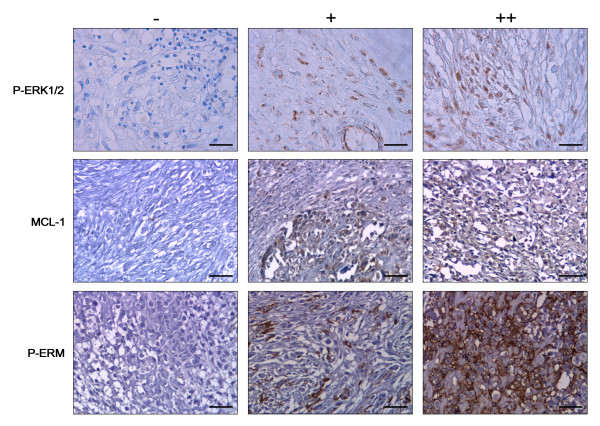
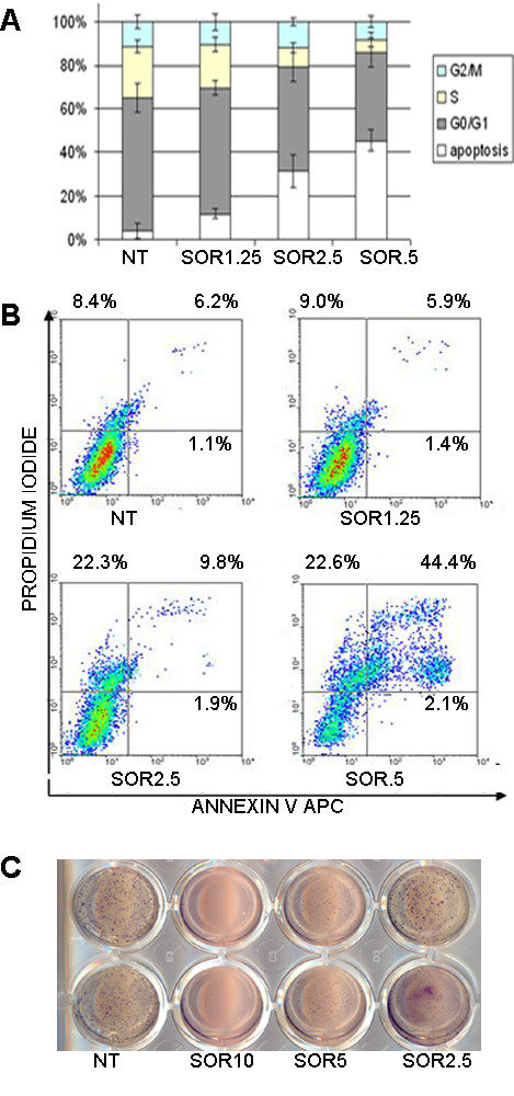
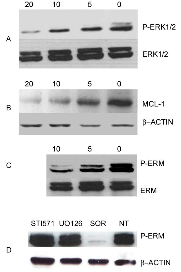
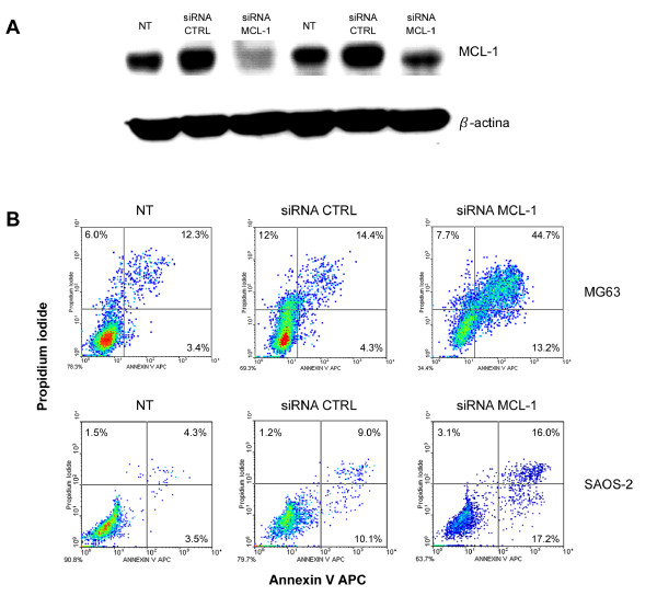
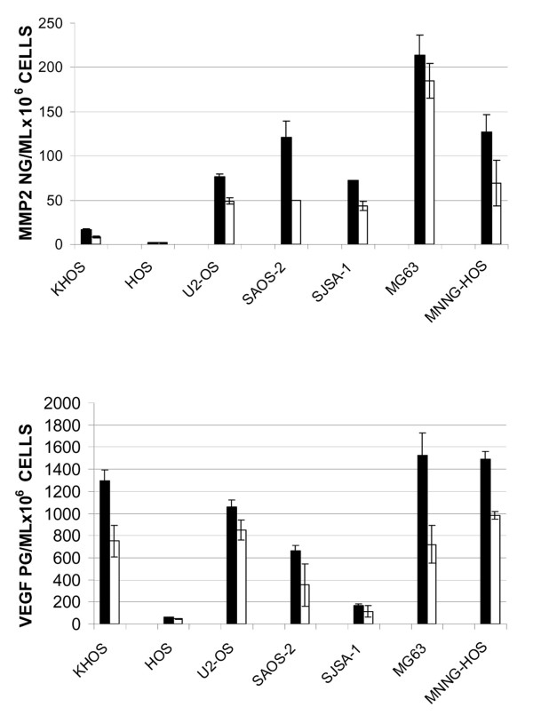
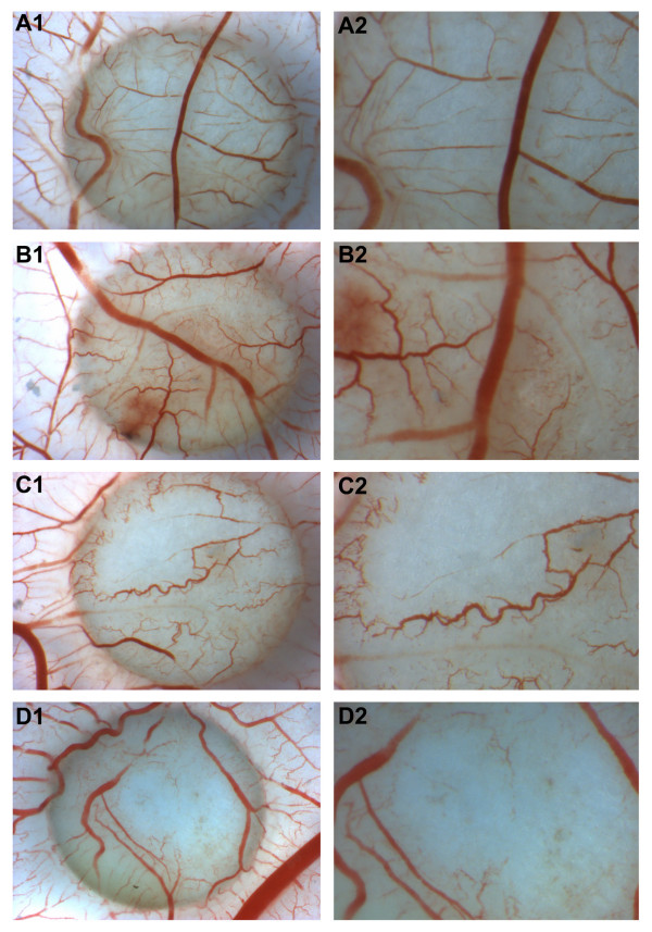
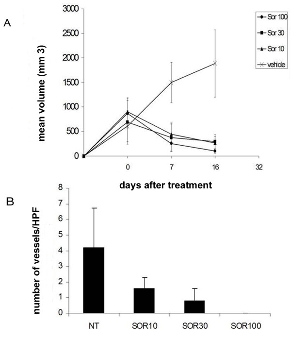
Results: We investigated phospho-ERK 1/2, MCL-1, and phospho-Ezrin/Radixin/Moesin (P-ERM) as potential therapeutic targets in OS. Activation of these pathways was shown by immunohistochemistry in about 70% of cases and in all OS cell lines analyzed. Mutational analysis revealed no activating mutations in KRAS whereas BRAF gene was found to be mutated in 4/30 OS samples from patients. Based on these results we tested the multi-kinase inhibitor sorafenib (BAY 43-9006) in preclinical models of OS. Sorafenib inhibited OS cell line proliferation, induced apoptosis and downregulated P-ERK1/2, MCL-1, and P-ERM in a dose-dependent manner. The dephosphorylation of ERM was not due to ERK inhibition. The downregulation of MCL-1 led to an increase in apoptosis in OS cell lines. In chick embryo chorioallantoic membranes, OS supernatants induced angiogenesis, which was blocked by sorafenib and it was also shown that sorafenib reduced VEGF and MMP2 production. In addition, sorafenib treatment dramatically reduced tumour volume of OS xenografts and lung metastasis in SCID mice.

Conclusion: In conclusion, ERK1/2, MCL-1 and ERM pathways are shown to be active in OS. Sorafenib is able to inhibit their signal transduction, both in vitro and in vivo, displaying anti-tumoural activity, anti-angiogenic effects, and reducing metastatic colony formation in lungs. These data support the testing of sorafenib as a potential therapeutic option in metastatic or relapsed OS patients unresponsive to standard treatments.


Figures

Figure 1
Figure 1
Representative immunostaining of osteosarcoma samples. Negative (-), positive (+) and strongly positive (++) samples for P-ERK1/2, MCL-1 and P-ERM expression.
Figure 2
Figure 2
Sorafenib induces apoptosis and inhibits anchorage independent cell growth of a representative OS cell line. DNA content (A) and apoptosis (B) analysis after 72 h treatments. Soft agar assays as described in "materials and methods" section (C).
Figure 3
Figure 3
Sorafenib induces down-regulation of P-ERK 1/2 (A), MCL-1 (B) and P-ERM (C) on U2OS. Effect on P-ERM is independent from PDGFR, C-KIT and ERK pathways (D). Cells were cultured for 24 hours with escalating doses (5, 10, 20 μM) of sorafenib, 10 μM STI571, 10 μM UO126 or left untreated. After protein extraction P-ERK 1/2 (A), MCL-1, (B), P-ERM (C and D) immunoblotting was carried out.
Figure 4
Figure 4
MCL-1 down-regulation induces apoptosis in OS cell lines. MCL-1 protein expression (A) and annexin V/PI staining (B) in MG63 and SAOS-2 after MCL-1 silencing with siRNA.
Figure 5
Figure 5
ELISA test for MMP2 and VEGF quantification on osteosarcoma cell line supernatants. The concentration of secreted MMP2 (a) and VEGF (B) on 48 h conditioned supernatants of untreated (Black bars) and 5 μM sorafenib treated (white bars) OS cells was shown per 106 cells as Mean +/- standard deviation (Y error bars) of 2 different experiments in triplicate. a, p < 0.05 versus untreated cells; b, p < 0.05 versus sorafenib.
Figure 6
Figure 6
Sorafenib inhibits angiogenesis in CAM mode. A. Culture medium (n = 4). A1: 10× magnification, A2: 20× magnification; B. Osteosarcoma cell line conditioned medium (n = 6). B1:10× magnification, B2: 20× magnification; C. Conditioned medium of sorafenib treated cells (n = 5). C1:10× magnification, C2: 20× magnification; D. Osteosarcoma conditioned medium plus sorafenib 1 μM (n = 7). D1:10× magnification, D2: 20× magnification.
Figure 7
Figure 7
Sorafenib activity in OS xenograft models. In three different experiments, one group of mice was injected s.c. into the right flank with U20S and one group of mice was injected e.v. with SJSA-1 cells. Five mice per group were orally treated daily with sorafenib (0-10-30-100 mg/kg/die) for 16 days as described in the Materials and Methods section. (A) Sorafenib reduces tumour volume OS xenografts in SCID mice. The graph indicates the mean tumour volume (mm3) measured using calipers at 0, 7 and 16 days of sorafenib treatment (Error bars: standard deviation). (B) Sorafenib reduces blood vessel number. The graph shows the mean number of patented blood vessel +/- standard deviation calculated in 10 fields at 40× magnification on Masson's trichromic staining.
Figure 8
Figure 8
Sorafenib reduces tumour mass in OS xenografts in SCID mice. Histological sections derived from sorafenib untreated (a) and treated (b) xenografts stained with Hematoxylin and Eosin show massive necrosis and reduced cellularity. Sorafenib reduces blood vessel number. 40× magnification on Masson's trichromic staining of sorafenib untreated (c) and treated (d) xenografts. Sorafenib reduces lung OS foci. Pictures show representative examples (10× magnification) of OS foci in lung sections of untreated (e) and sorafenib-treated mice (f).

Similar articles

Cited by

References

    1. Ferrari S, Smeland S, Mercuri M, Bertoni F, Longhi A, Ruggieri P, Alvegard TA, Picci P, Capanna R, Bernini G, Müller C, Tienghi A, Wiebe T, Comandone A, Böhling T, Del Prever AB, Brosjö O, Bacci G, Saeter G. Italian and Scandinavian Sarcoma Groups. Neoadjuvant chemotherapy with high-dose Ifosfamide, high-dose methotrexate, cisplatin, and doxorubicin for patients with localized osteosarcoma of the extremity: a joint study by the Italian and Scandinavian Sarcoma Groups. J Clin Oncol. 2005;23:8845–52. doi: 10.1200/JCO.2004.00.5785. - DOI - PubMed
    1. Ferrari S, Briccoli A, Mercuri M, Bertoni F, Picci P, Tienghi A, Del Prever AB, Fagioli F, Comandone A, Bacci G. Postrelapse survival in osteosarcoma of the extremities: prognostic factors for long-term survival. J Clin Oncol. 2003;21:710–15. doi: 10.1200/JCO.2003.03.141. - DOI - PubMed
    1. Ozaki T, Flege S, Kevric M, Lindner N, Maas R, Delling G, Schwarz R, von Hochstetter AR, Salzer-Kuntschik M, Berdel WE, Jürgens H, Exner GU, Reichardt P, Mayer-Steinacker R, Ewerbeck V, Kotz R, Winkelmann W, Bielack SS. Osteosarcoma of the pelvis: experience of the Cooperative Osteosarcoma Study Group. J Clin Oncol. 2003;21:334–41. doi: 10.1200/JCO.2003.01.142. - DOI - PubMed
    1. Fagioli F, Aglietta M, Tienghi A, Ferrari S, Brach del Prever A, Vassallo E, Palmero A, Biasin E, Bacci G, Picci P, Madon E. High-dose chemotherapy in the treatment of relapsed osteosarcoma: an Italian sarcoma group study. J Clin Oncol. 2002;20:2150–6. doi: 10.1200/JCO.2002.08.081. - DOI - PubMed
    1. Serra M, Pasello M, Manara MC, Scotlandi K, Ferrari S, Bertoni F, Mercuri M, Alvegard TA, Picci P, Bacci G, Smeland S. May P-glycoprotein status be used to stratify high-grade osteosarcoma patients? Results from the Italian/Scandinavian Sarcoma Group 1 treatment protocol. Int J Oncol. 2006;29:1459–68. - PubMed

Publication types

MeSH terms