. 2009 Dec 10:10:594.
doi: 10.1186/1471-2164-10-594.

Insight into microRNA regulation by analyzing the characteristics of their targets in humans

Affiliations

Insight into microRNA regulation by analyzing the characteristics of their targets in humans

Zihua Hu. BMC Genomics. .

Abstract

Background: microRNAs (miRNAs) are believed to regulate their targets through posttranscriptional gene regulation and have the potential to silence gene expression via multiple mechanisms. Despite previous advances on miRNA regulation of gene expression, little has been investigated from a genome scale.

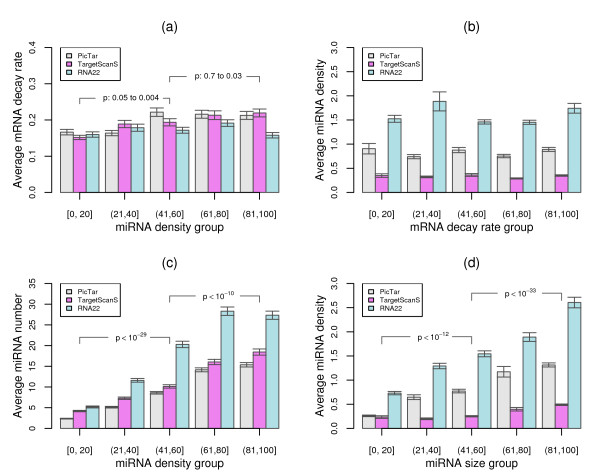
Results: To gain new insight into miRNA regulation in humans, we used large scale data and carried out a series of studies to compare various features of miRNA target genes to that of non-miRNA target genes. We observed significant differences between miRNA and non-miRNA target genes for a number of characteristics, including higher and broader mRNA expression, faster mRNA decay rate, longer protein half-life, and longer gene structures. Based on these features and by analyzing their relationships we found that miRNA target genes, other than having miRNA repression, were most likely under more complex regulation than non-miRNA target genes, which was evidenced by their higher and broader gene expression but longer gene structures. Our results of higher and broader gene expression but fast mRNA decay rates also provide evidence that miRNA dampening of the output of preexisting transcripts facilitates a more rapid and robust transition to new expression programs. This could be achieved by enhancing mRNA degradation through an additive effect from multiple miRNA targeting.

Conclusion: Genome-scale analysis on the nature of miRNA target genes has revealed a general mechanism for miRNA regulation of human gene expression. The results of this study also indicate that miRNA target genes, other than having miRNA repression, are under more complex gene regulation than non-miRNA target genes. These findings provide novel insight into miRNA regulation of human gene expression.


Figures

Figure 1
Figure 1
Expression and expression breadth differences between miRNA and non-miRNA target genes. (a) Distribution of median expression values from 79 human tissues and p-values for the difference between miRNA and non-miRNA target genes. Error bars indicate standard errors. (b) Distribution of the fraction of miRNA and non-miRNA target genes restrictedly expressed in certain number of tissues; p-values: statistical differences of median fractions between miRNA and non-miRNA target genes in each bin by Wilcoxon signed rank tests. The results from PicTar predicted miRNA target genes are shown.
Figure 2
Figure 2
Comparison of mRNA decay rate and protein stability between miRNA and non-miRNA target genes. (a) miRNA target genes on average have higher mRNA decay rates than non-miRNA target genes. (b) miRNA target genes on average have longer protein half-lives than non-miRNA target genes. Target genes predicted by three algorithms of PicTar, TargetScanS, and RNA22 are shown. Error bars indicate standard errors. p: p-values from one-side Wilcoxon rank sum tests for the median value differences between miRNA and non-miRNA target genes.
Figure 3
Figure 3
Correlation between mRNA expression and decay rate. (a) Spearman's rank correlation rho between gene expression from each of the 79 human tissues and mRNA decay rates, and corresponding p-values (-log10(p-values)) for the correlation coefficients. (b) Distribution of the average mRNA decay rates, which were obtained from comparing gene expression in each of the 79 human tissues, for 5 mRNA expression groups with increasing expression values from the group [1-20] to the group (81,100]. (c) Distribution of the average mRNA expression values in the 79 human tissues for 5 mRNA decay rate groups with increasing mRNA decay rate from the group [1-20] to the group (81,100]. Exp: mRNA expression; MDR: mRNA decay rate.
Figure 4
Figure 4
Correlation between gene expression and protein stability. (a) Spearman's rank correlation rho between gene expression from each of the 79 human tissues and protein stability, and corresponding p-values (-log10(p-values)) for the correlation coefficients. (b) Distribution of the average protein stability indices, which were obtained from comparing gene expression in each of the 79 human tissues, for 5 mRNA expression groups with increasing expression values from the group [1,20] to the group (81,100]. (c) Distribution of the average mRNA expression values in the 79 human tissues for 5 protein stability groups with increasing protein stability index from the group [1,20] to the group (81,100]. Exp: mRNA expression; PSI: protein stability. Index.
Figure 5
Figure 5
Relationship between the number of miRNA binding sites and mRNA decay rate. (a) Variation of mRNA decay rates for 5 miRNA binding site groups with increasing number of binding sites. The degree of the increasing trend for mRNA decay rates along with the increasing number of miRNA binding sites was estimated by Wilcoxon rank sum tests and depicted in p-values. (b) The expected variation of mRNA decay rates from both permutation tests and shuffled 3'UTR sequences for 5 miRNA binding site groups with increasing number of binding sites. (c) Variation of miRNA binding site number for 5 mRNA decay rate groups with increasing decay rates. The degree of the increasing trend for miRNA binding site number along with the increasing mRNA decay rates was estimated by Wilcoxon rank sum tests and depicted in p-values. (d) The expected variation of miRNA binding site number from both permutation tests and shuffled 3'UTR sequences for 5 mRNA decay rate groups with increasing decay rates.
Figure 6
Figure 6
Relationship between miRNA density, mRNA decay rate, and the number of miRNA binding sites. (a) Variation of mRNA decay rates for 5 miRNA density groups with increasing miRNA density. The degree of the increasing trend for mRNA decay rates along with the increasing miRNA density was estimated by Wilcoxon rank sum tests and depicted in p-values. (b) Variation of miRNA density for 5 mRNA decay rate groups with increasing decay rate values. (c) Variation of miRNA binding site number for 5 miRNA density groups with increasing density values. The degree of the increasing trend for miRNA binding site number along with the increasing miRNA density was estimated by Wilcoxon rank sum tests and depicted in p-values. (d) Variation of miRNA density for 5 miRNA binding site groups with increasing number of binding sites. The degree of the increasing trend for miRNA density along with the increasing number of miRNA binding sites was estimated by Wilcoxon rank sum tests and depicted in p-values.
Figure 7
Figure 7
Relationship between the length of 3'UTR and mRNA decay rate. Variation of mRNA decay rates of different 3'UTR length groups for miRNA target genes with different miRNA target sites predicted from PicTar (a), TargetScanS (b), and RNA22 (c). The median length of each 3'UTR length group is shown in the legend boxes. The degree of the mRNA decay rate differences between the low, median, and high 3'UTR length groups in each miRNA binding site groups was estimated by Wilcoxon rank sum tests and the significant ones are depicted in p-values.

Similar articles

Cited by

References

    1. Lee RC, Feinbaum RL, Ambros V. The C. elegans heterochronic gene lin-4 encodes small RNAs with antisense complementarity to lin-14. Cell. 1993;75(5):843–854. doi: 10.1016/0092-8674(93)90529-Y. - DOI - PubMed
    1. Wightman B, Ha I, Ruvkun G. Posttranscriptional regulation of the heterochronic gene lin-14 by lin-4 mediates temporal pattern formation in C. elegans. Cell. 1993;75(5):855–862. doi: 10.1016/0092-8674(93)90530-4. - DOI - PubMed
    1. Kloosterman WP, Plasterk RH. The diverse functions of microRNAs in animal development and disease. Developmental cell. 2006;11(4):441–450. doi: 10.1016/j.devcel.2006.09.009. - DOI - PubMed
    1. Voinnet O. Origin, biogenesis, and activity of plant microRNAs. Cell. 2009;136(4):669–687. doi: 10.1016/j.cell.2009.01.046. - DOI - PubMed
    1. Sarnow P, Jopling CL, Norman KL, Schutz S, Wehner KA. MicroRNAs: expression, avoidance and subversion by vertebrate viruses. Nat Rev Microbiol. 2006;4(9):651–659. doi: 10.1038/nrmicro1473. - DOI - PubMed

Publication types

LinkOut - more resources